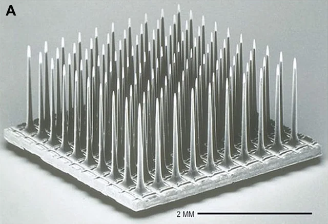
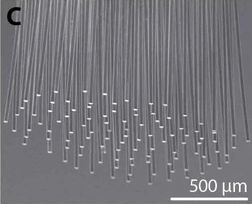
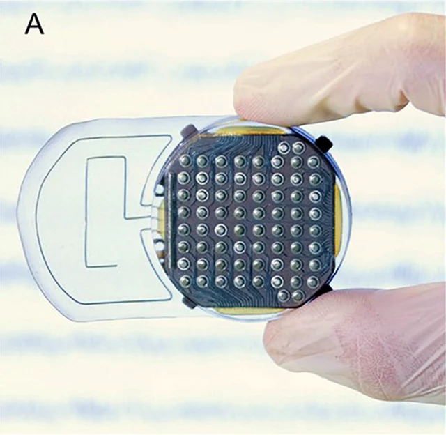
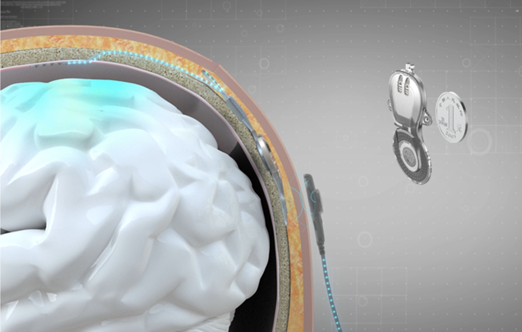
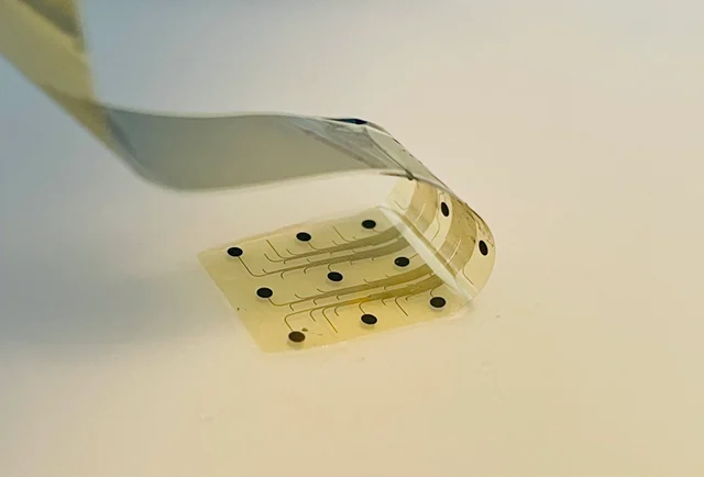
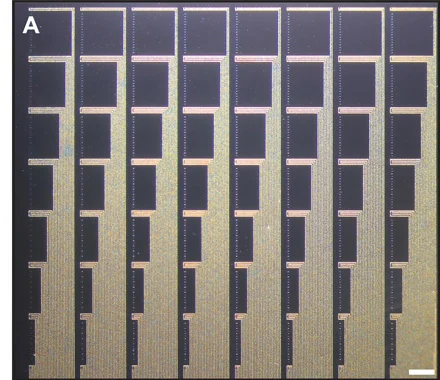
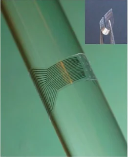
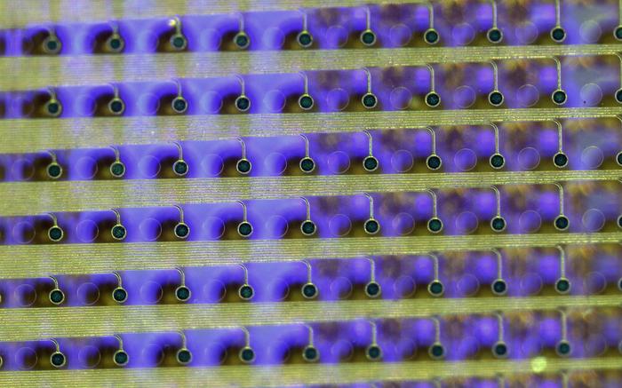
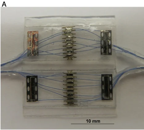
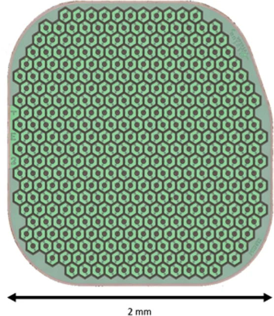
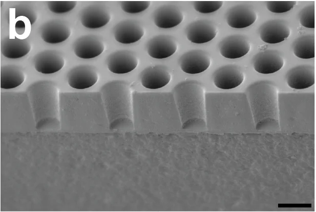
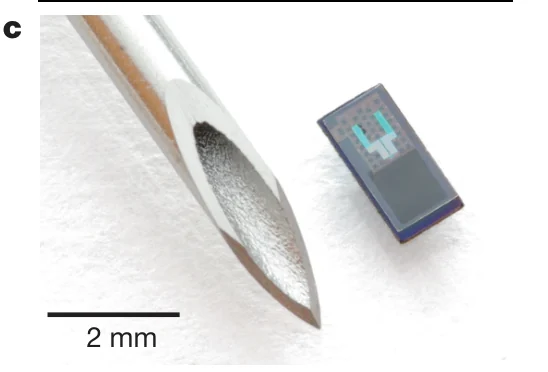
View the interface → Electron micrograph C3? Utah electrode array, scanning electron micrograph. 2 mm scale retained.
Utah arrays - Parallel structural variants
Utah Microelectrode Array (UEA) Silicon penetrating arrays with 400 µm electrode pitch. Separates the Utah research family from the 100-electrode, 96-connected NeuroPort configuration and records conflicting manufacturer counts.
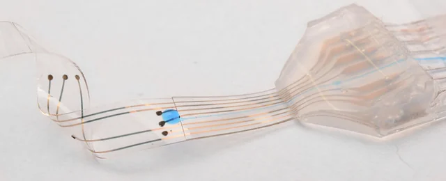
The slanted array targets different depths in peripheral nerves. It does not supersede the standard cortical Utah array.
3D model available Intracortical Human evidence
Photo credit Fernandez et al., Frontiers in Neuroengineering (2014), Fig. 1A · Frontiers · Figure 1A · CC BY 3.0 . Photographic panel cropped from the original figure; resized and re-encoded. Original scale bars and annotations retained.
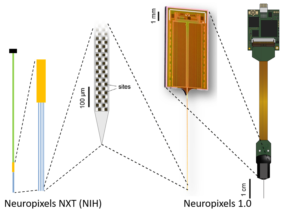
Past versions and parallel branches (1) View the interface → Source figure L2R The official NXT page illustrates the project’s four-shank architecture and enlarged recording-site region beside Neuropixels 1.0.
Neuropixels - Generations and parallel branches
Neuropixels NXT / NP3.0 prototype Official 2026 prototype report: 1,536-channel four-shank NXT, planned NP3.0 name, up to 912 channels mapped to one shank. Access and 2027 sale are forecasts, not completed deliveries.
NXT / NP3.0 is the latest announced generation, still a prototype. Opto, Ultra and NHP solve different problems and remain separate branches.
3D model available Intracortical Preclinical evidence
Photo credit Neuropixels Central / Johns Hopkins, NXT project page · Neuropixels Central / Johns Hopkins, NXT project page · Source copyright retained; educational reference reproduced at site owner request . Original source image preserved intact, byte for byte; all panels, scale bars and annotations retained.
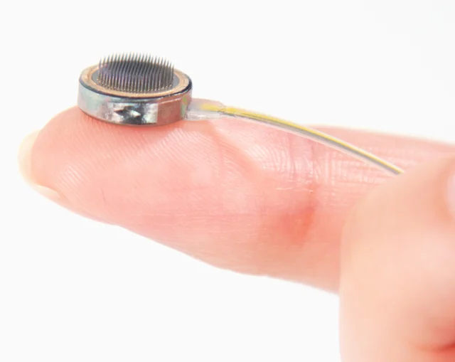
Past versions and parallel branches (6) View the interface → Photograph C3R Connexus cortical module on a fingertip, showing its penetrating microelectrodes.
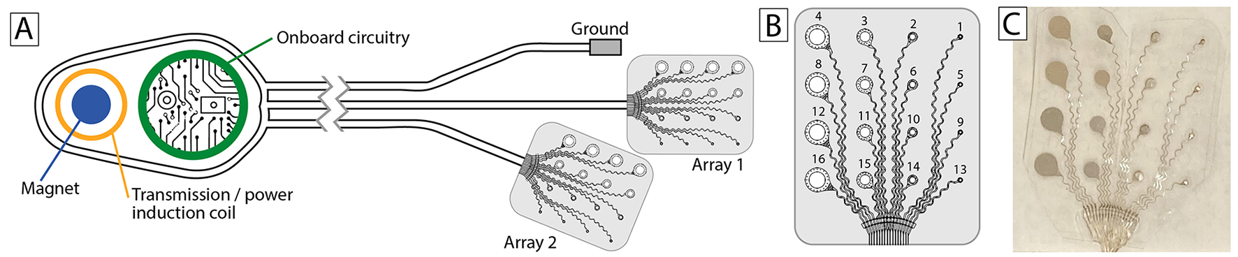
Paradromics Connexus (cortical module) Paradromics’ 421-electrode intracortical module, with a source-documented 3D reference model and updated Connect-One early feasibility study context.
3D model available Intracortical Human evidence
Photo credit Paradromics, Inc. — official press kit · Paradromics · official press kit · Official press kit — editorial use with credit . Editorial crop centered on the cortical module; resized and re-encoded. No hardware retouching.
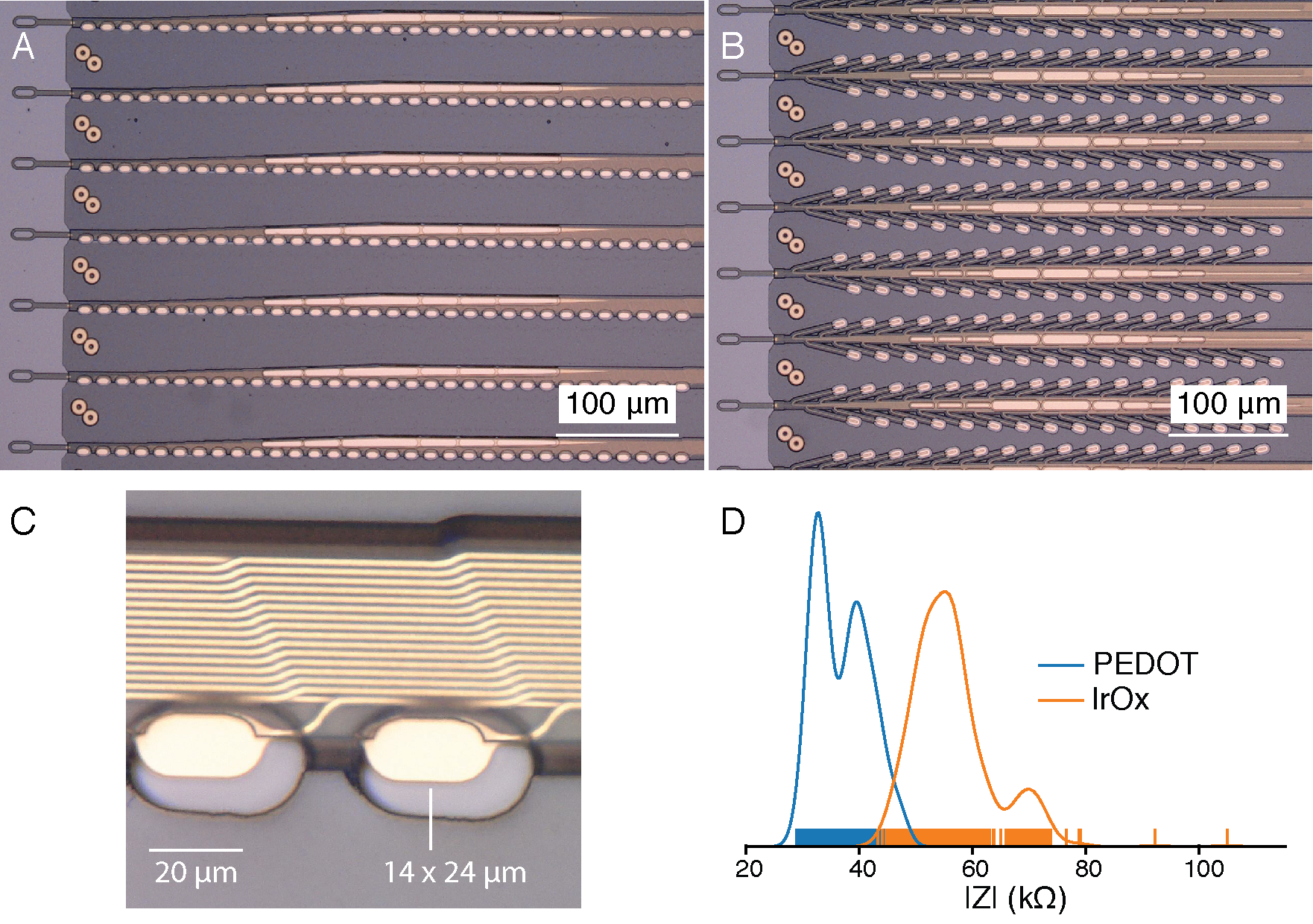
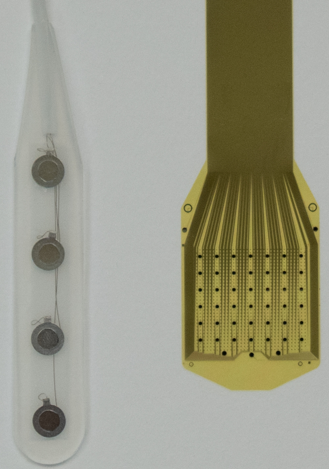
View the interface → Source figure · 2 views C3R Neuralink’s 2019 research thread architecture: two polymer probe designs, electrode contacts and impedance measurements. Historical reference, not the current N1 implant.
Neuralink N1 (flexible-thread implant) Neuralink’s fully implanted, wireless intracortical BCI, with a 3D reference for the documented 2024 N1 configuration: 64 flexible threads and 1,024 sites.
3D model available Intracortical Human evidence
Photo credit Elon Musk and Neuralink, 2019 research white paper, published in JMIR 21(10):e16194 · Neuralink · 2019 research white paper · CC BY-ND 4.0 . Complete figure recovered at its native embedded resolution from the 2019 white paper. Transparency composited on the paper’s white background; no cropping, upscaling, retouching or panel removal. The recovered PNG is copied intact for both display and enlargement.
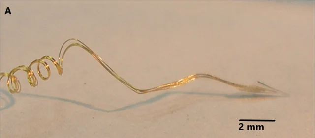
View the interface → Optical micrograph ?3R Neurotrophic cone-electrode assembly photographed under magnification; 2 mm scale retained.
Neurotrophic Electrode (Kennedy cone electrode) A biologically integrated intracortical electrode (hollow cone with microwires) designed to encourage neurite ingrowth for long-term single-unit recording in humans.
Intracortical Human evidence
Photo credit Gearing and Kennedy, Frontiers in Human Neuroscience (2020), Fig. 1A · Frontiers · Figure 1A · CC BY 4.0 . Photographic panel cropped from the original figure; resized and re-encoded. Original scale bars and annotations retained.
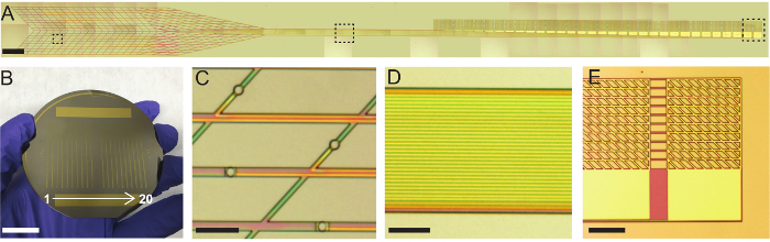
View the interface → Source figure L2R Fabricated mesh electronics and their connection pads, shown across the complete photographic Figure 3.
Syringe-injectable mesh electronics Sub-micrometre-thick, centimetre-scale mesh electronics that unfold after injection through a needle as small as 100 µm. Harvard, 2015, in mouse brain.
Intracortical Preclinical evidence
Photo credit Schuhmann et al., Journal of Visualized Experiments (2018), Figure 3 · JoVE · complete Figure 3 · CC BY-NC-ND 3.0 . Complete source image retained byte-for-byte, including every panel and annotation. No cropping, re-encoding, recoloring or synthetic enhancement.
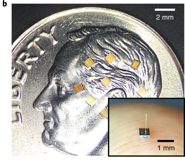
View the interface → Photograph · 2 views L2R Actual neurograins, with 2 mm scene scale and 1 mm inset scale.
Neurograins (wireless microimplant network) Distributed RF-powered neurograins: 650 × 650 × 250 µm chiplets, 1-kHz/8-bit recording, 48-node acute rat ensemble. Clear low-noise recordings came from a fraction of those channels; 770 is a projected network capacity.
3D model available Other Preclinical evidence
Photo credit Lee et al., Nature Electronics (2021), Fig. 1b · Lee et al., Nature Electronics (2021), Fig. 1b · Source copyright retained; owner-requested noncommercial reference . Source panel mechanically extracted; source annotations and scale bars retained. Resized and re-encoded.
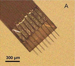
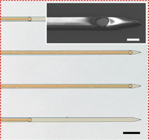
View the interface → Optical micrograph ?2R Michigan carbon-fiber flex array, pictured in Huan et al. (2021): a related 2 × 8, 132µm-pitch configuration with sharpened PEDOT:pTS-coated tips, not the exact 2020 bare-carbon implant.
Carbon fiber electrode arrays (University of Michigan) Michigan carbon-fiber arrays: 2015 PEG or silicon-assisted cortical recording and 2020 flex arrays for separate-session unit and dopamine sensing. Coatings, pitch, supports and cohorts differ.
3D model available Intracortical Preclinical evidence
Photo credit Huan et al., Journal of Neural Engineering 18 (2021), Figure 1A. DOI: 10.1088/1741-2552/ac3dd7 · Original source · photographic panel · CC BY 4.0 . Photographic panel cropped from the original figure; resized and re-encoded. Original scale bars and annotations retained.
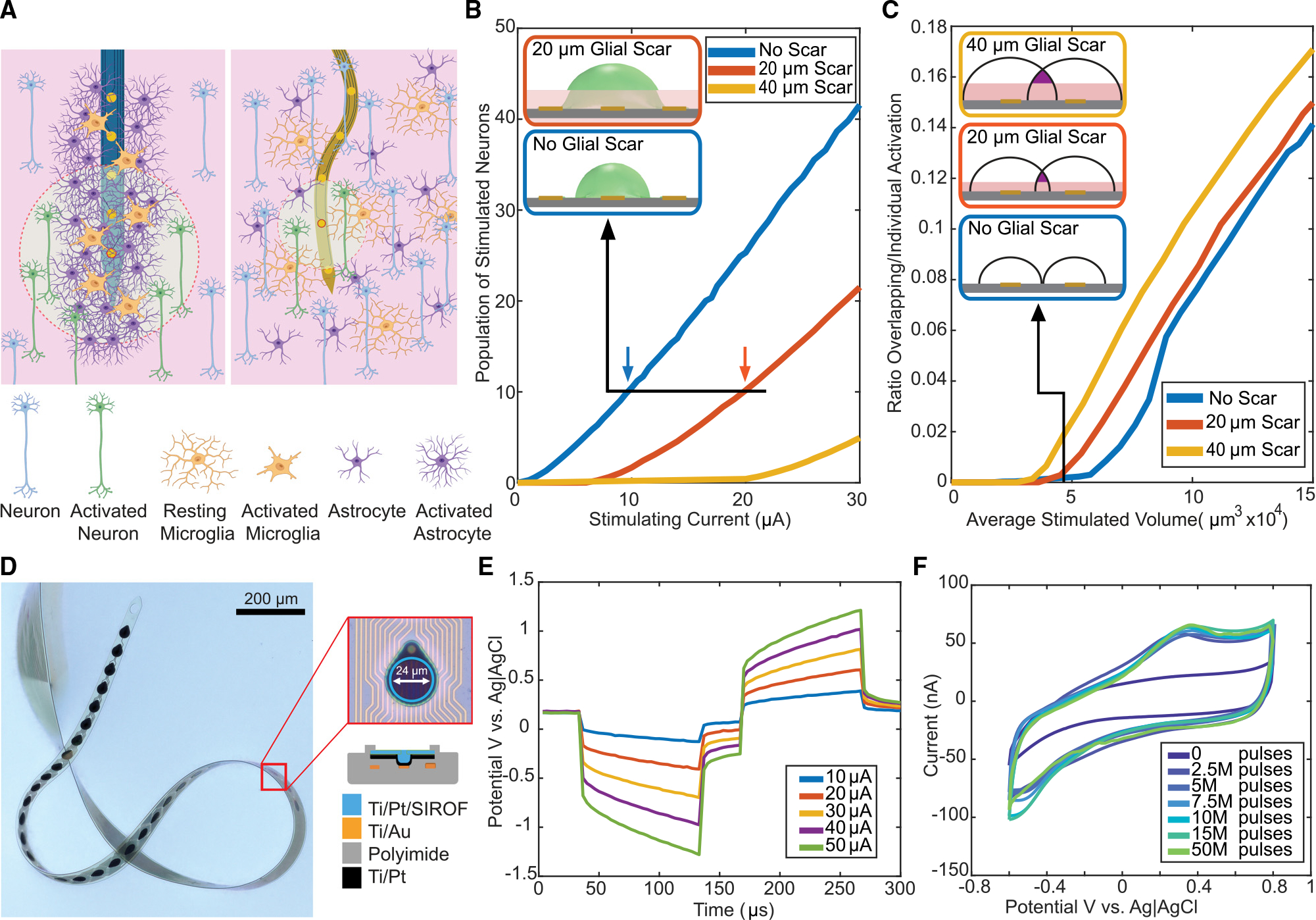
View the interface → Source figure L2R Panel D shows the real ultraflexible StimNET in water, with an enlarged 24 µm contact and the polyimide cap-ring stack; the full source figure is retained.
Nanoelectronic threads - Recording lineage and stimulation branch
StimNET ultraflexible stimulation thread Rice's 2023 32-contact polyimide stimulation/recording thread: 1 µm shank, reinforced sputtered-iridium-oxide contacts and separate bench and chronic mouse stimulation evidence.
StimNET is the latest documented branch here, designed for stimulation. Modular NET remains a distinct high-channel-count recording system.
Intracortical Preclinical evidence
Photo credit Lycke et al., Cell Reports (2023), Figure 1 · Lycke et al., Cell Reports (2023), Figure 1 · CC BY-NC-ND 4.0; reproduced complete and unmodified . Original source image preserved intact, byte for byte; all panels, scale bars and annotations retained.
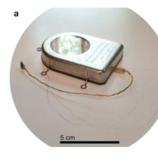
Past versions and parallel branches (2) View the interface → Photograph · 3 views L2R 2013 Brown sealed implant assembly with 5 cm source scale.
Brown Wireless Neural Interface A subcutaneous titanium-housed system that streams 100 channels of broadband cortical data wirelessly and charges through the skin. Brown University, tested in moving primates, 2013.
Intracortical Preclinical evidence
Photo credit Borton et al., Journal of Neural Engineering (2013), Fig. 2a · Borton et al., Journal of Neural Engineering (2013), Fig. 2a · Source copyright retained; owner-requested noncommercial reference . Source panel mechanically extracted; source annotations and scale bars retained. Resized and re-encoded.
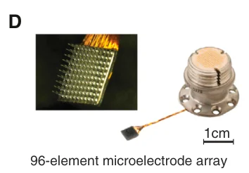
View the interface → Source figure · 2 views L2R Implanted 96-electrode array and pedestal used with the external Brown 2014 neurosensor; original Figure 1D.
Brown Wireless Neurosensor External Brown2014 neurosensor: 96 neural channels plus auxiliary paths, 20 kS/s per channel, 46.1-g headstage and over48-hour reported runtime. Battery chemistry wording conflict retained; not a fully implanted package.
Intracortical Preclinical evidence
Photo credit Yin et al., Neuron (2014), Fig. 1D; EPFL author-hosted PDF · Yin et al., Neuron (2014), Fig. 1D; EPFL author-hosted PDF · Source copyright retained; owner-requested noncommercial reference . Source figure resized and re-encoded; all panels, annotations and scale bars retained.
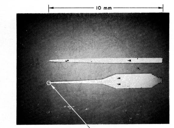
View the interface → Optical micrograph · 2 views ?1R Wise 1970 double-electrode probe; original 10 mm length dimension retained.
Wise Silicon Microprobe The first multielectrode microprobe made with integrated-circuit fabrication: gold electrodes on a silicon carrier, patterned by photoengraving. Wise, Angell and Starr, 1970. The root of the Michigan probe line.
Intracortical Research evidence
Photo credit Wise, Angell and Starr, IEEE TBME (1970), Fig. 8 overview · Wise, Angell and Starr, IEEE TBME (1970), Fig. 8 overview · CC BY 4.0 . Source panel mechanically extracted; source annotations and scale bars retained. Resized and re-encoded.
DEVICE IMAGE Photograph pending verification ?2R
Image unavailable · device record retained Neuralink Blindsight Neuralink's visual prosthesis, which would stimulate visual cortex directly so that people without working eyes or optic nerves might perceive images. FDA breakthrough device designation in September 2024. Human results are not confirmed in the sources used here.
Intracortical Preclinical evidence
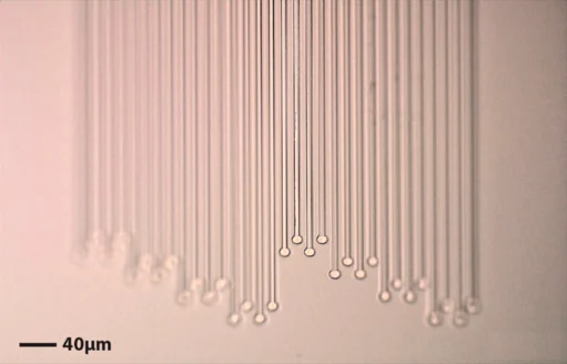
View the interface → Optical micrograph ?2R NeuroRoots electrode tendrils and contacts under optical microscopy. Scale bar: 40 µm.
NeuroRoots (independent flexible electrode tendrils) Independent 7 µm wide, 1.5 µm thick electrode tendrils. The 2024 paper reports cerebellar single-unit recordings and at least seven weeks of rat recordings without repositioning.
Intracortical Preclinical evidence
Photo credit NeuroRoots study authors, APL Bioengineering (2024), Figure 1B, CC BY-NC 4.0 · Original source · photographic panel · CC BY-NC 4.0 . Photographic panel cropped from the original figure; resized and re-encoded. Original scale bars and annotations retained.
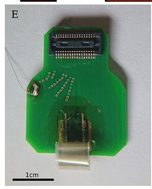
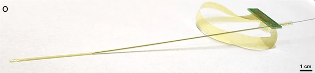
View the interface → Photograph · 3 views ?2R Fabricated sewn-polymer electrode threads epoxied and wirebonded to a connector PCB; threads curl from parylene stress. Source 1 cm scale retained.
Sewn Polymer Electrode Array A 2019 preprint system with 64 individually inserted polyimide-platinum threads, 16 µm shanks and one site per thread. Rat recordings exposed implant durability and tissue-response limits.
Intracortical Preclinical evidence
Photo credit Hanson et al., bioRxiv (2019), Fig. 2E · Hanson et al., bioRxiv (2019), Fig. 2E · CC BY-NC 4.0 . Source figure panel extracted from the original preprint PDF; scale bars and annotations retained. Rendered and re-encoded.
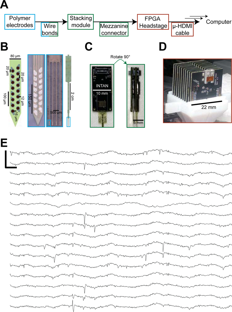
View the interface → Source figure ?2R Complete Figure 1 documents the implanted polymer shanks separately from the headstage electronics.
Modular polymer probe system (UCSF and Lawrence Livermore) Polyimide arrays with 16 contacts per shank and stackable 64-channel modules, supporting up to 1,024 recording channels in freely behaving rats. Neuron, 2019.
Intracortical Preclinical evidence
Photo credit Chung et al., Neuron (2019), Figure 1 · Chung et al., Neuron (2019), Figure 1 · Source copyright retained; site-owner-requested noncommercial scholarly reference . Original source image copied intact, byte for byte; labels, panels and scale bars retained.
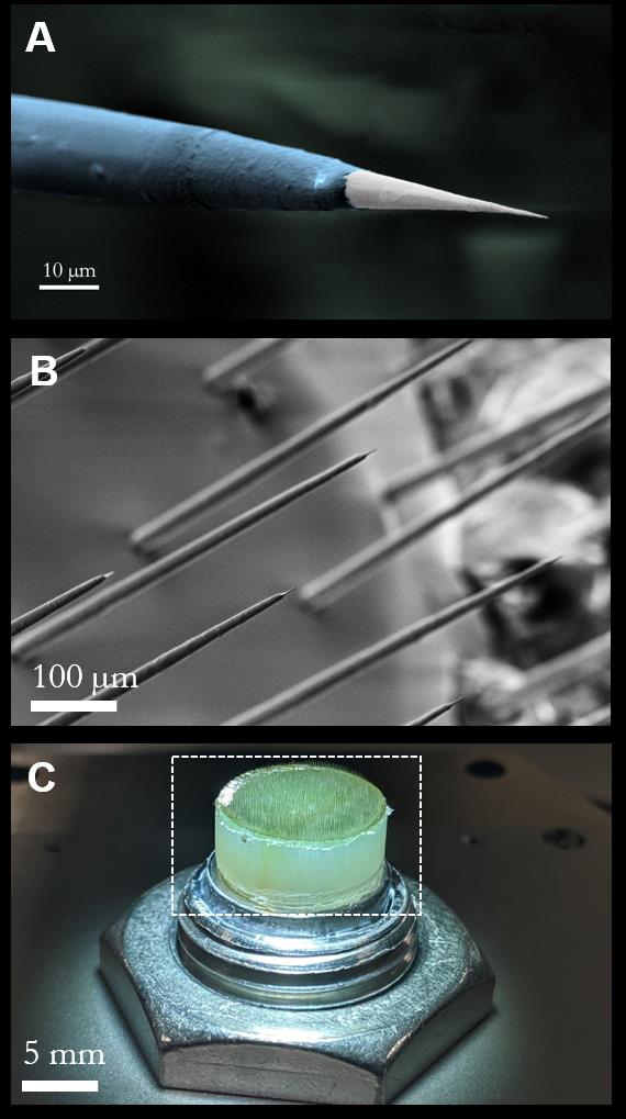
View the interface → Source figure C2R Argo microwire tip (A), electrode-tip array (B), and assembled bundle (C); the original scale bars are retained.
Argo microwire-CMOS recording system Paradromics/Caeleste's 2021 acute research system: a 65,536-pixel CMOS readout bonded to disordered PtIr wires, with 1,300-wire rat spiking and over 30,000 connected sheep surface channels kept separate.
Other Preclinical evidence
Photo credit Sahasrabuddhe et al., bioRxiv preprint (2020), Figure 2 · bioRxiv · complete Figure 2 · CC BY-NC-ND 4.0 . Complete source image retained byte-for-byte, including every panel and annotation. No cropping, re-encoding, recoloring or synthetic enhancement.
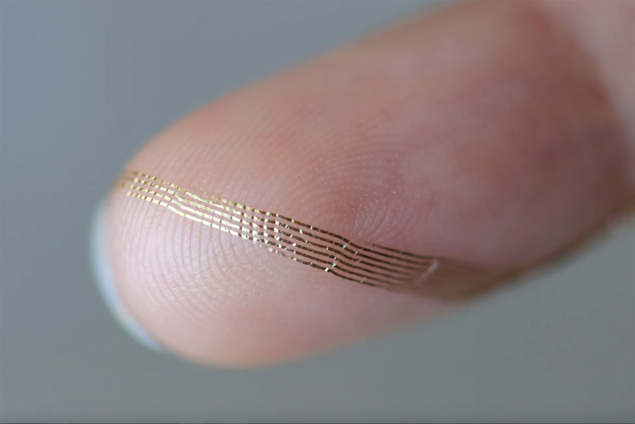
View the interface → Photograph · 3 views C3R Axoft Fleuron neural probe photographed on a fingertip. Fine traces and exposed site rows are visible.
Axoft Fleuron ultrasoft neural probe Ultrasoft 128-channel depth probe from Axoft, tested for about 20 minutes in tissue due for resection in a first-in-human study in 2025. Preprint and company claims; not a chronic implant.
Intracortical Human evidence
Photo credit Axoft, via Harvard Office of Technology Development (2025) · Axoft, via Harvard Office of Technology Development (2025) · Copyright retained by source; owner-requested noncommercial reference . Original source image preserved byte for byte; all embedded panels, annotations and scale bars retained.
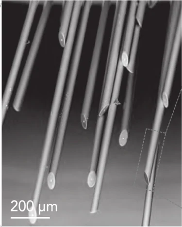
View the interface → Electron micrograph ?2R The tissue-facing end of a CHIME glass-insulated microwire bundle, imaged by scanning electron microscopy. Scale bar: 200 µm.
CHIME glass-gold microwire CMOS interface, 2020 CHIME's glass-insulated gold wires and electroplated contacts coupled to MEA1k or camera-derived CMOS. Demonstrated bundles and connected pixels are separate from327,680 amplifier capacity; chronic validation remains open.
Intracortical Preclinical evidence
Photo credit Kollo et al., Frontiers in Neuroscience (2020), Fig. 2C detail, CC BY 4.0 · Frontiers · original figure · CC BY 4.0 . Select bottom central SEM inset of Figure 2C, retaining original 200 µm scale.
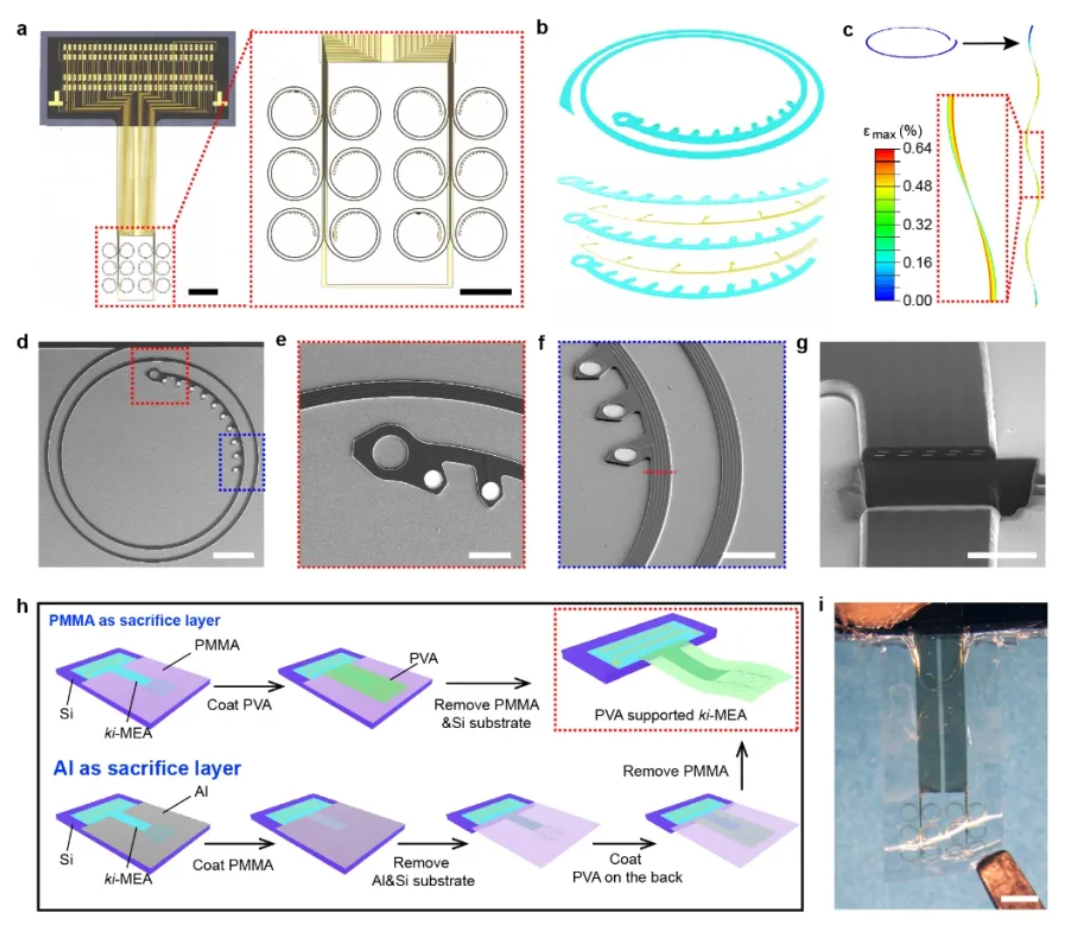
View the interface → Source figure · 2 views ?2R Complete figure of the 120-channel research ki-MEA, including the planar kirigami pattern, microscopy, 3D mechanics and fabrication panels.
CIBR kirigami microelectrode array (ki-MEA) for primate brain Stretchable penetrating array whose electrode wires are cut like kirigami and form helical springs once implanted. 120-channel rodent and 256-channel macaque versions, 250 days of recording with over 240 neurons per session, about 1000 channels across arrays and a reach-decoding R2 of 0.79. Fang Ying's lab at CIBR Beijing (Nature Electronics, Feb 2026).
Intracortical Preclinical evidence
Photo credit Fang et al., Nature Electronics (2026), Fig. 2, via CIBR · Fang et al., Nature Electronics (2026), Fig. 2, via CIBR · Copyright retained by source; owner-requested noncommercial reference . Original source image preserved byte for byte; all embedded panels, annotations and scale bars retained.
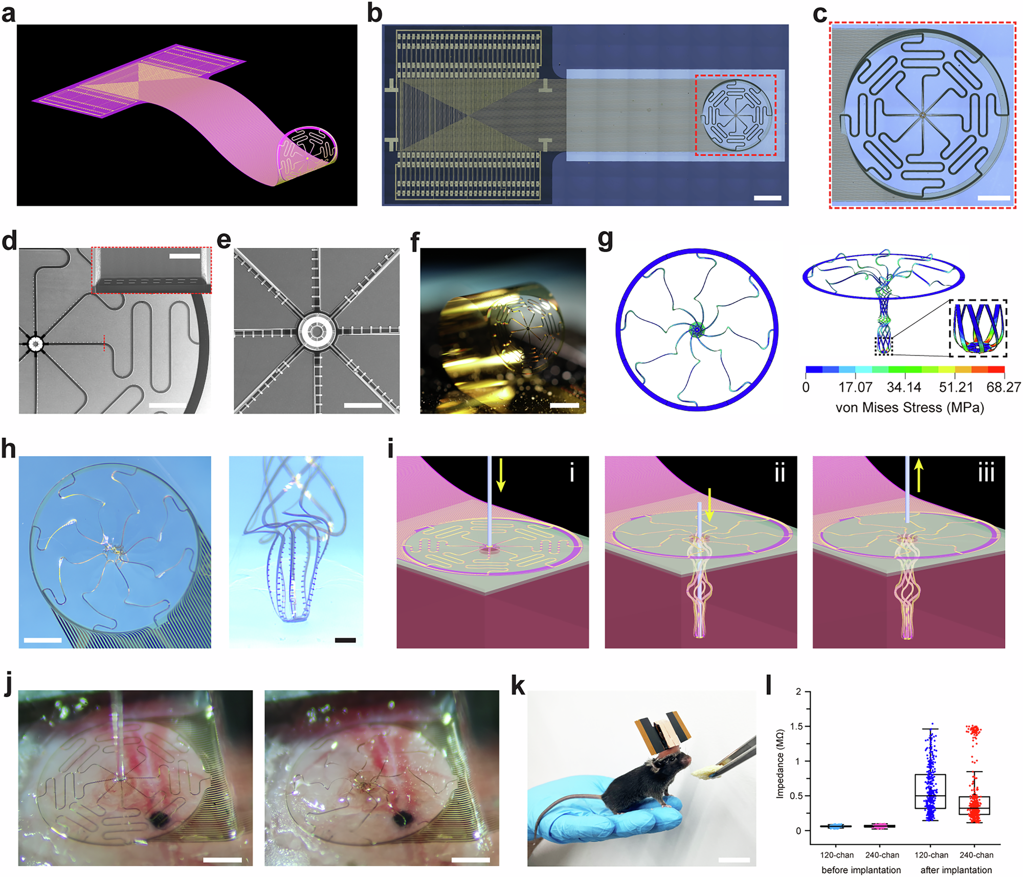
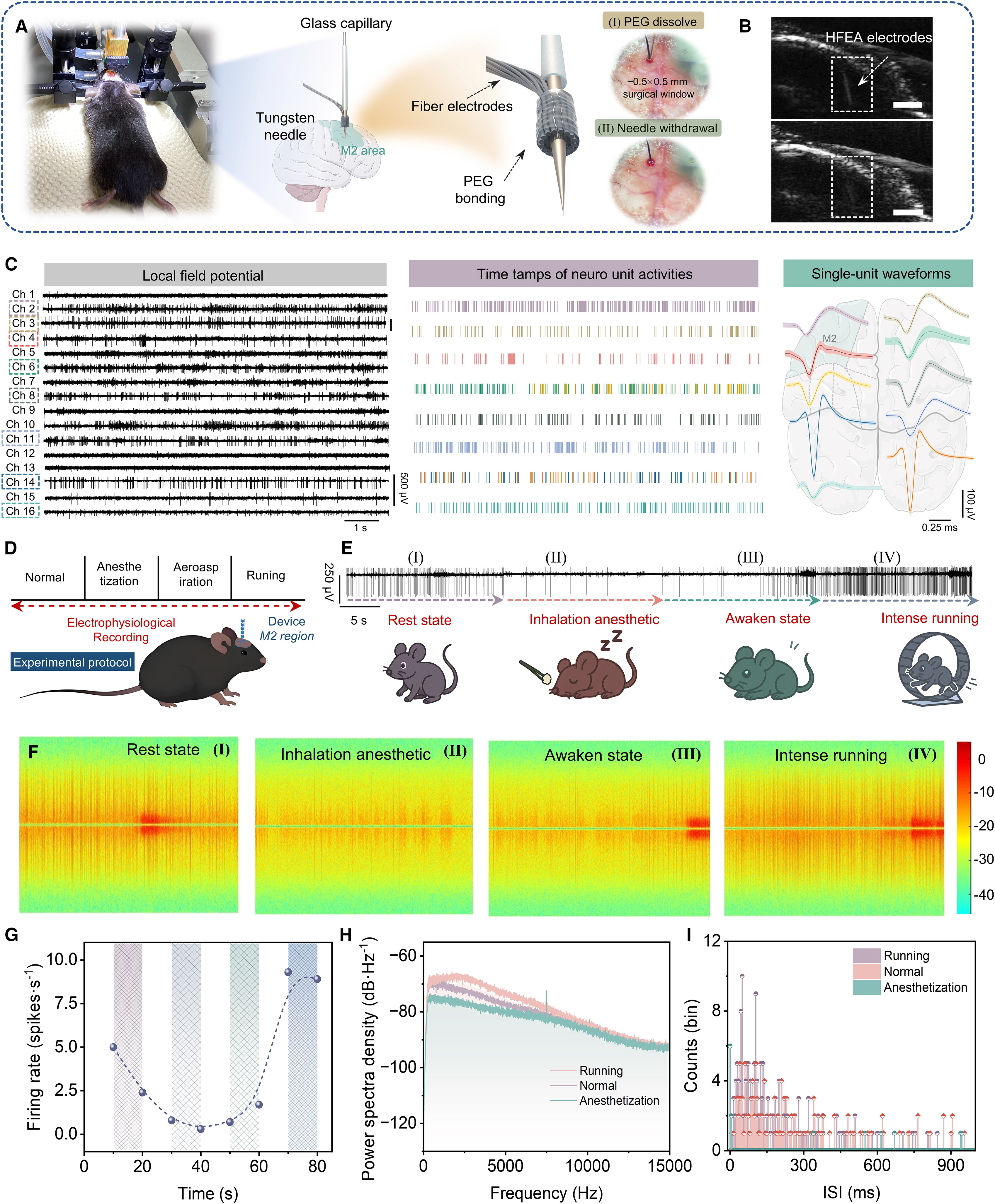
View the interface → Source figure ?2R Figure 1 of the paper: the umbrella-type array and probe in micrographs and photographs (b to f, h, scale bars as published), photographs of the implanted array on mouse cortex (j) and a mouse with the implant (k, scale bar as published), with renderings, a simulation and impedance data in the remaining panels.
CIBR umbrella-type stretchable electrode array (USEs probe) 120- and 240-channel ultrathin (about 3 micrometer) polyimide and gold array of eight stretchable serpentine filaments joined to a central anchor, implanted in one pass on a 190 micrometer shuttle. Gao Lei's lab, Chinese Institute for Brain Research Beijing; same neuronal ensembles tracked in head-fixed mice for 80 weeks (Nature Communications 2026).
Intracortical Preclinical evidence
Photo credit Chinese Institute for Brain Research (CIBR) group and collaborators, Nature Communications (2026), Figure 1 · Nature Communications · complete Figure 1 · CC BY-NC-ND 4.0 . Complete source image retained byte-for-byte, including every panel and annotation. No cropping, re-encoding, recoloring or synthetic enhancement.
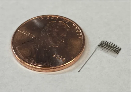
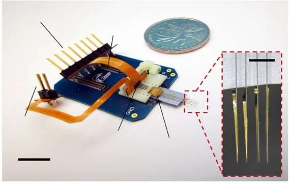
View the interface → Photograph ?2R The released flexible neurochemical and recording probe, beside a US penny for scale.
CMU Multimodal Neurochemical Probe Parylene C probe with 16 recording contacts and two electrically actuated chemical-release sites. Acute rat proof of concept, with finite drug loading, stimulation artifacts and chronic-use work still open.
Intracortical Preclinical evidence
Photo credit Malekoshoaraie et al., Microsystems & Nanoengineering (2024), Fig. 2a, CC BY 4.0 · Publisher · original figure · CC BY 4.0 . Select only photo panel 2a; no recoloring, geometry alteration or invented background. Penny retained as scale.
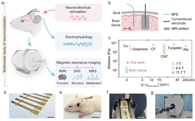
View the interface → Source figure L2R Figure 1 of the paper: photographs of the fibre electrode array (d), a rat carrying the implant (e), the animal in the MRI bed (f) and the scanner (g), with schematic and plot panels a to c. Scale bars as published.
Fudan MRI-compatible PEDOT:PSS fiber electrode (MFE) Wet-spun PEDOT:PSS fibre about 15 micrometers across with PDMS insulation and a magnetic susceptibility of -9.23 ppm matched to brain tissue, giving little to no artifact at 11.7 T. 11.9 kOhm at 1 kHz for a 20 micrometer fibre, charge injection limit 14.3 mC/cm2, used to stimulate and record in rats during multimodal MRI. Fudan University (National Science Review 2026).
Intracortical Preclinical evidence
Photo credit Fudan University group, National Science Review (2026), Figure 1 · National Science Review · complete Figure 1 (CC BY 4.0) · CC BY 4.0 . Complete source image retained byte-for-byte as served by PubMed Central (661 px wide rendition). No cropping, re-encoding, recoloring or synthetic enhancement.
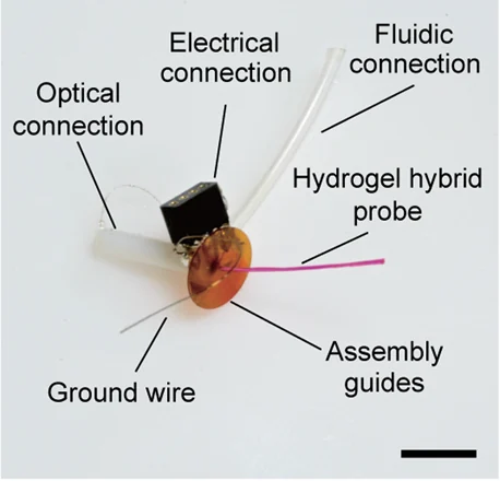
View the interface → Photograph L2R The hydrogel-hybrid fiber assembly, with a 1 cm scale bar. The hydrogel is dyed pink for visibility in the source photograph.
Hydrogel-hybrid multifunctional fiber probe (MIT) Thermally drawn optical, electrical and fluidic fibers integrated in a soft hydrogel matrix. Dry insertion and hydrated compliance, with six-month mouse recordings and a connectorization limit that prevents independent readout of every microwire.
Intracortical Preclinical evidence
Photo credit Park et al., Nature Communications (2021), Fig. 1e, CC BY 4.0 · Publisher · original figure · CC BY 4.0 . Select source photo panel 1e only, with original labels and scale; no hardware editing.
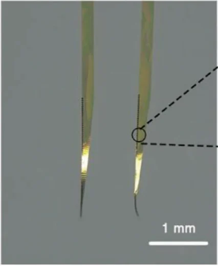
View the interface → Optical micrograph · 2 views ?2R Ultraflexible MERF recording shanks; 1 mm scale retained.
MERF 128-channel ultraflexible polyimide array (CEBSIT) Two-shank, 128-channel, 1 micrometer thick polyimide array from Zhao Zhengtao's group at CEBSIT (Chinese Academy of Sciences) with Li Chengyu's group at Lingang Laboratory. Chronic single-unit recording in macaque visual and motor cortex for up to eight months, plus a cursor-control demonstration.
Intracortical Preclinical evidence
Photo credit Tian et al., Advanced Science (2023), Figure 1b · Advanced Science · Figure 1b · CC BY 4.0 . Photographic panel rendered directly from the published PDF in its original colors, cropped and re-encoded. Original scales retained; no retouching or synthetic enhancement.
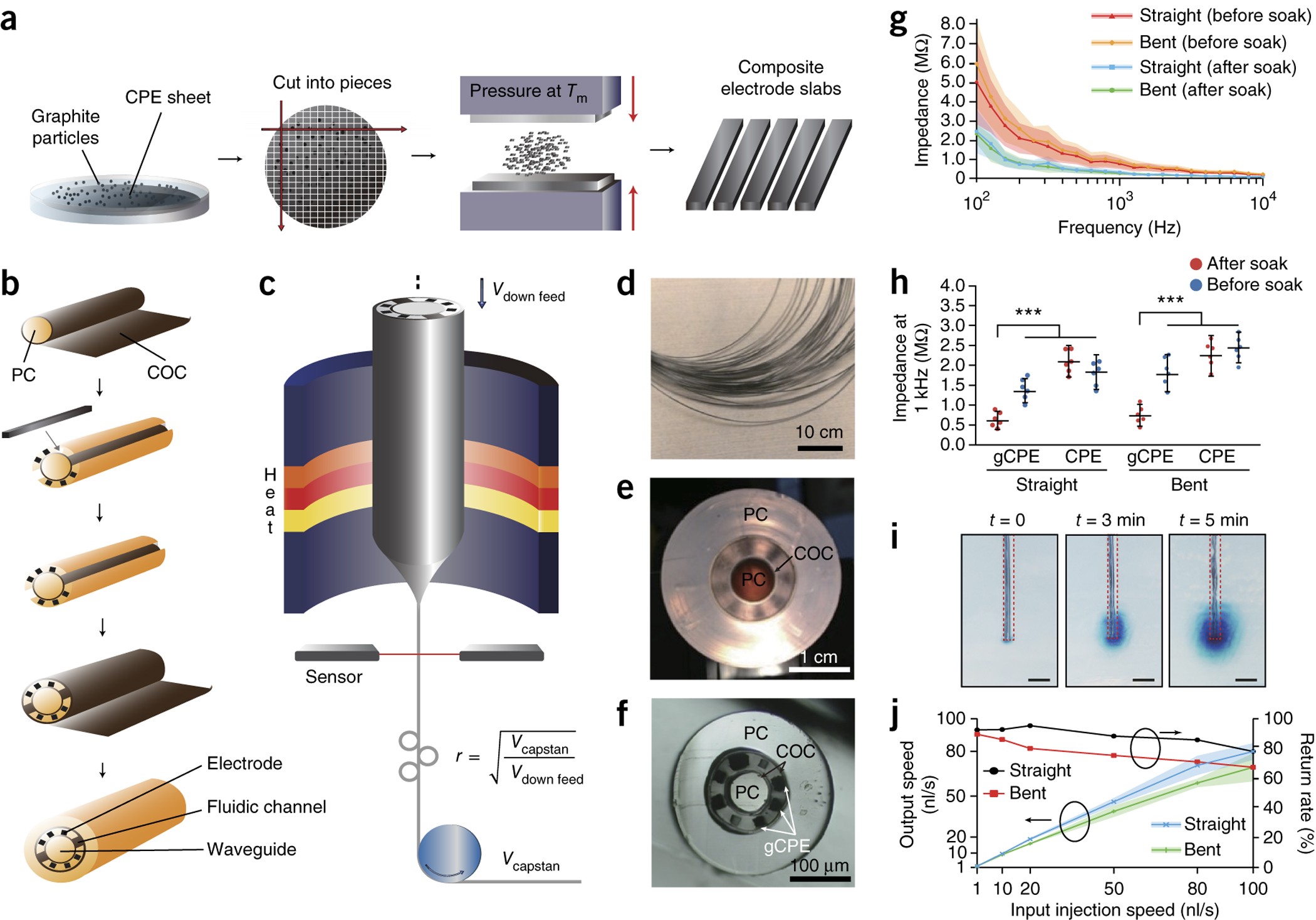
View the interface → Source figure · 2 views L2R The primary 2017 figure shows the actual drawn fiber cross-section with graphite-doped conductive-polymer electrodes, waveguide and fluidic channels.
MIT all-polymer drawn fibers - Published hardware lineage
MIT graphite-polymer multifunctional fiber, 2017 Graphite-doped polymer electrodes, one optical waveguide and two fluidic channels within a drawn fiber. The selected experimental section was 200 micrometers despite a less-than-200 claim elsewhere in the paper.
The 2017 graphite-polymer design develops the 2015 all-polymer fiber. Tin-wire and hydrogel-hybrid devices are different architectures, not hidden revisions.
Intracortical Preclinical evidence
Photo credit Park et al., Nature Neuroscience (2017), Figure 1 · Park et al., Nature Neuroscience (2017), Figure 1 · Source copyright retained; educational reference reproduced at site owner request . Original source image preserved intact, byte for byte; all panels, scale bars and annotations retained.
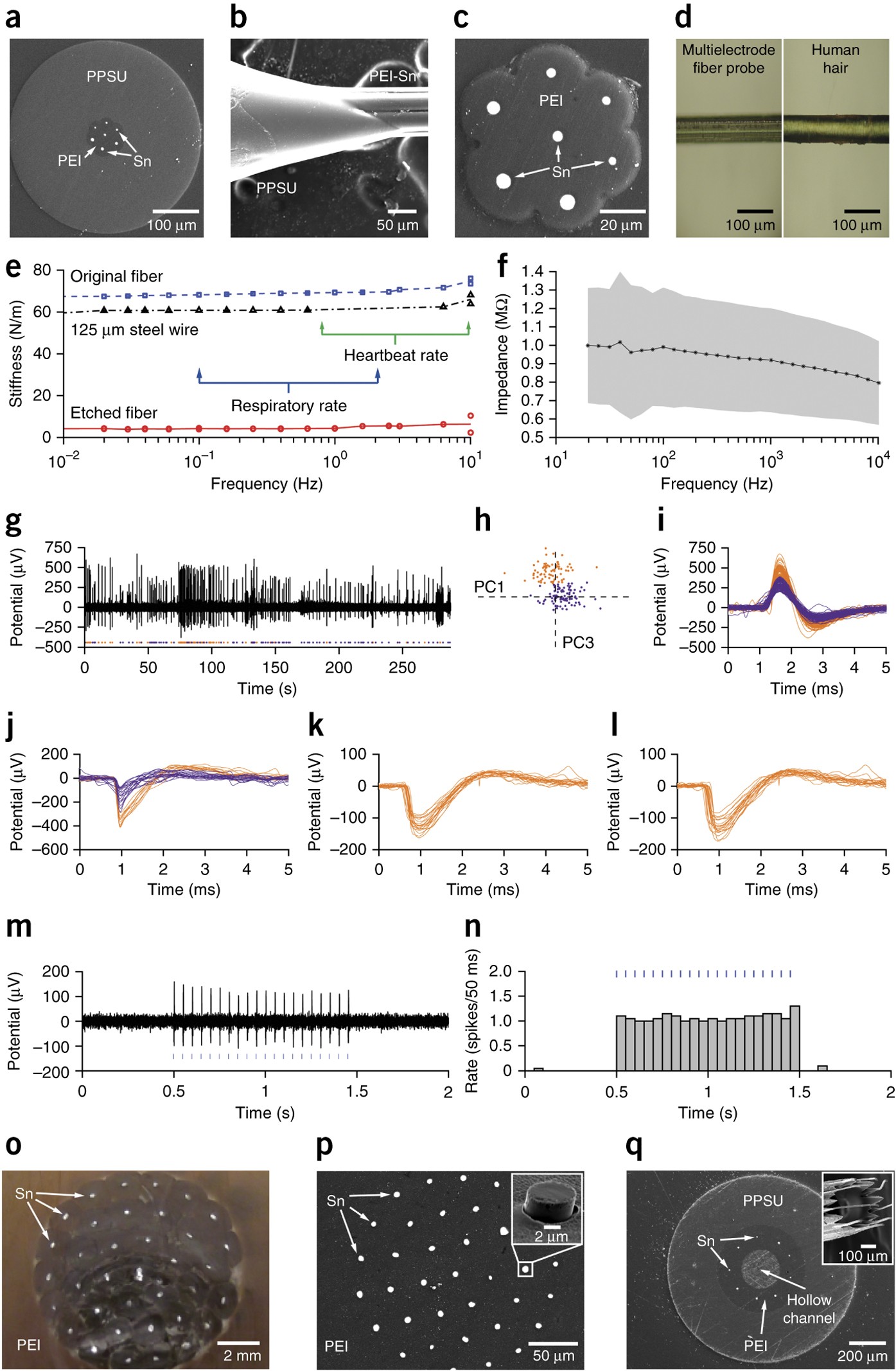
Past versions and parallel branches (1) View the interface → Source figure L2R Figure 3 specifically documents the tin/PEI multielectrode design, including its exposed cross-section and thin etched structure; the source’s additional architectures remain separately labeled.
MIT thermally drawn tin multielectrode fiber, 2015 Tin electrodes in a polymer fiber, with sacrificial-cladding removal reducing the demonstrated cross-section from 416 to 85 micrometers. Recording-only architecture is distinct from the all-polymer optical/fluidic fiber.
Intracortical Preclinical evidence
Photo credit Canales et al., Nature Biotechnology (2015), Figure 3 · Canales et al., Nature Biotechnology (2015), Figure 3 · Source copyright retained; educational reference reproduced at site owner request . Original source image preserved intact, byte for byte; all panels, scale bars and annotations retained.
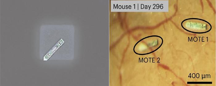
View the interface → Optical micrograph L2R MOTE on a salt crystal (left), and implanted MOTEs in mouse cortex at day 296 (right); 400 µm scale retained.
MOTE optical tetherless recorder, 2025 Subnanolitre electrical recorder with AlGaAs optical power and PPM uplink. Mouse evidence uses a cranial window and head fixation; day-365 LFP is weakened and averaged.
Intracortical Preclinical evidence
Photo credit KAIST · KAIST · original research press image · Institutional press-image permission · credit retained . Complete source image retained byte-for-byte, including every panel and annotation. No cropping, re-encoding, recoloring or synthetic enhancement.
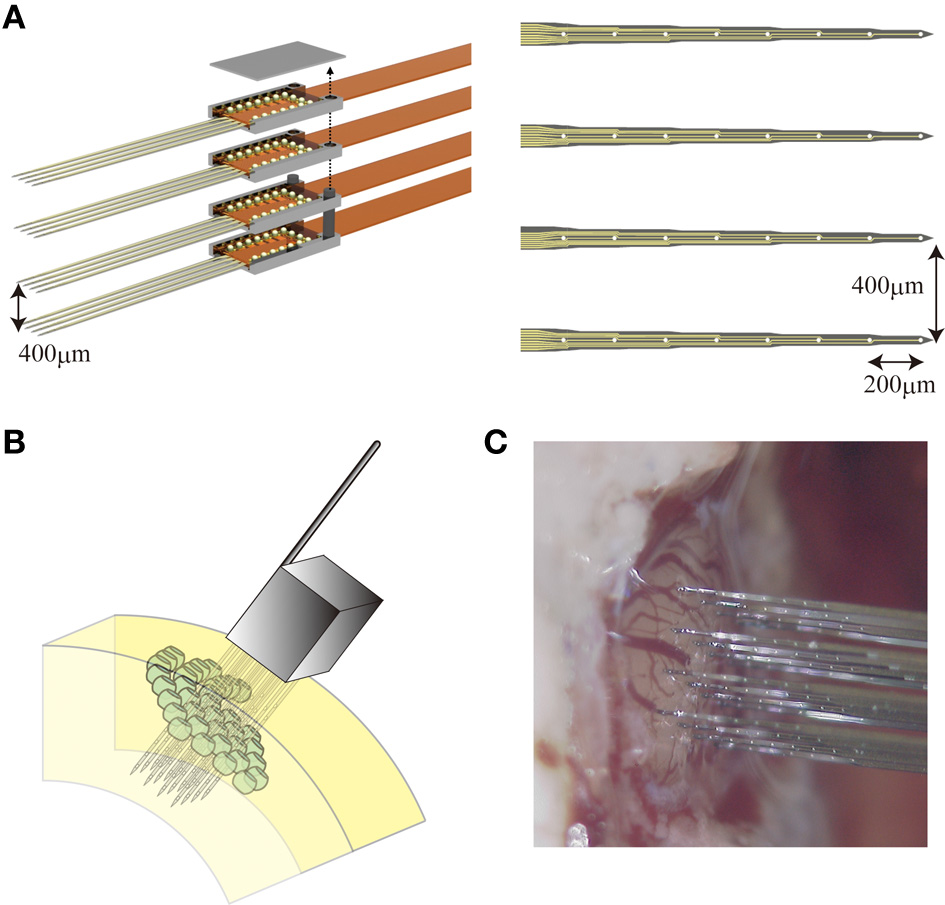
View the interface → Source figure ?1R NeuroNexus planar silicon probes assembled into a 3D recording array; research photograph and geometry drawings.
NeuroNexus silicon probe and array families NeuroNexus Technologies' configurable microelectrode probe and array families for extracellular recording and stimulation, including silicon shank arrays and Intan-compatible X-Series probes, with supporting electrophysiology systems.
Intracortical Research evidence
Photo credit Riera, Goto and Kawashima, Frontiers in Neural Circuits (2014), Figure 1; assembly credited to NeuroNexus · Frontiers in Neural Circuits · Figure 1 · CC BY 3.0 . Original complete research figure copied byte for byte to local storage; no crop, resizing or re-encoding. Original panels, annotations and scale bars retained.
View the interface → Optical micrograph ?2R The recording ends of Neurotassel filaments under optical microscopy. Scale bar: 50 µm; electron-micrograph inset: 10 µm.
Neurotassel elastocapillary filament arrays China-secondary coverage: 2019 polyimide filaments self-assembled in dissolvable PEG; 16-channel chronic mouse evidence is separate from 128-1,024-channel fabrication and preliminary recording claims.
Intracortical Preclinical evidence
Photo credit Guan, Wang, Gu et al., Science Advances (2019), Fig. 1B, CC BY-NC 4.0 · Original source · physical interface · CC BY-NC 4.0 . Photographic panel cropped from the original figure; resized and re-encoded. Original annotations and scale bars retained where present.
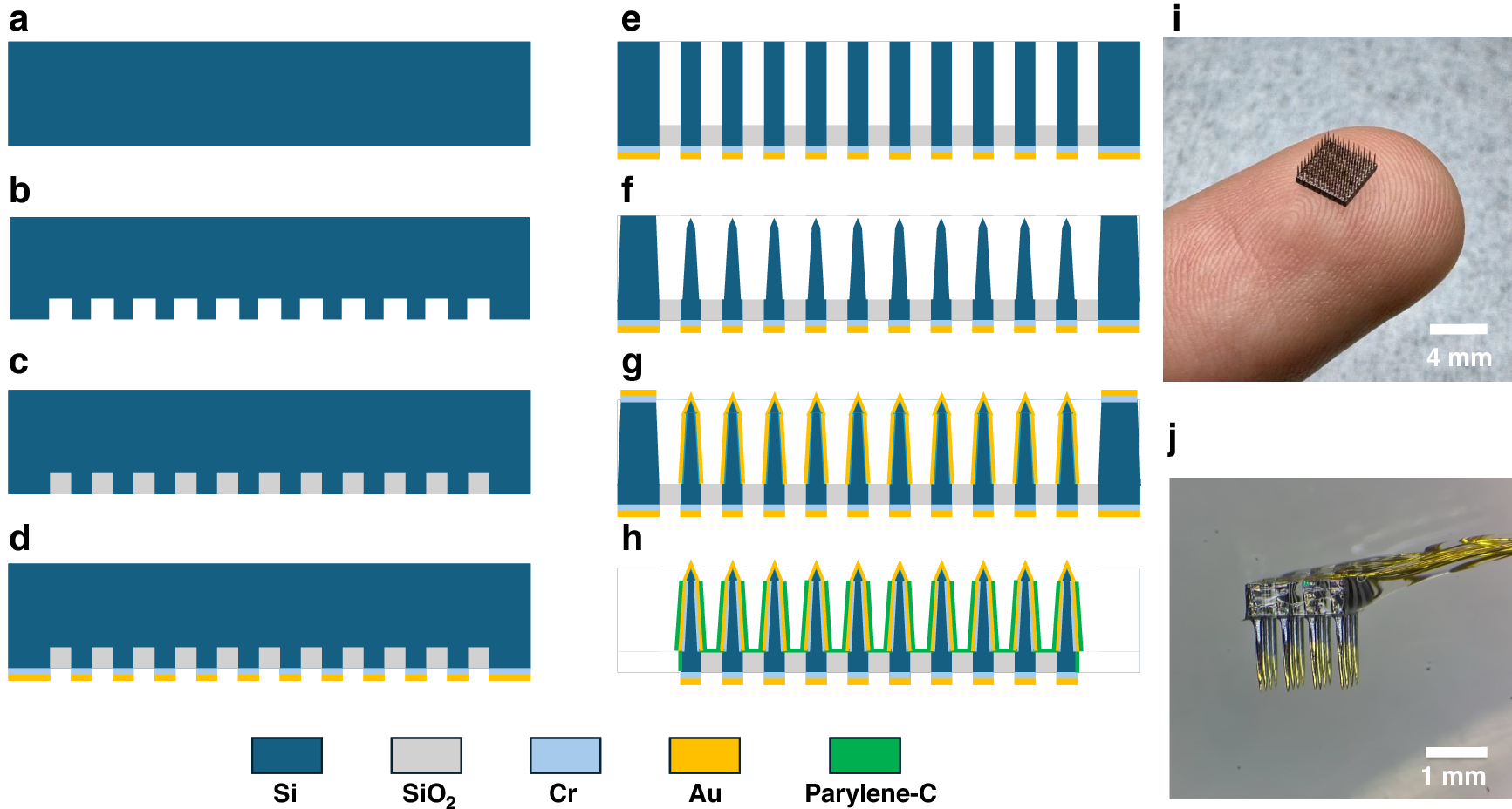
View the interface → Source figure ?2R Fabrication process (a to h, schematics) and photographs of the completed array (i, 4 mm scale) and a packaged array (j, 1 mm scale), shown across the complete Figure 1.
Semi CAS silicon microneedle array with local de-insulation 10 x 10 bulk-silicon microneedle array, Utah-style, with tips exposed by masking in PDMS before Parylene-C deposition instead of etching. Tip exposure about 50 um, platinum black sites at 33.2 kOhm at 1 kHz, 15 of 16 channels recording in rat with mean spike SNR 12.63. Pei Weihua's group, Institute of Semiconductors, CAS.
Intracortical Preclinical evidence
Photo credit Pei group, Institute of Semiconductors CAS, Microsystems & Nanoengineering (2025), Figure 1 · Microsystems & Nanoengineering · complete Figure 1 · CC BY-NC-ND 4.0 . Complete source image retained byte-for-byte, including every panel and annotation. No cropping, re-encoding, recoloring or synthetic enhancement.
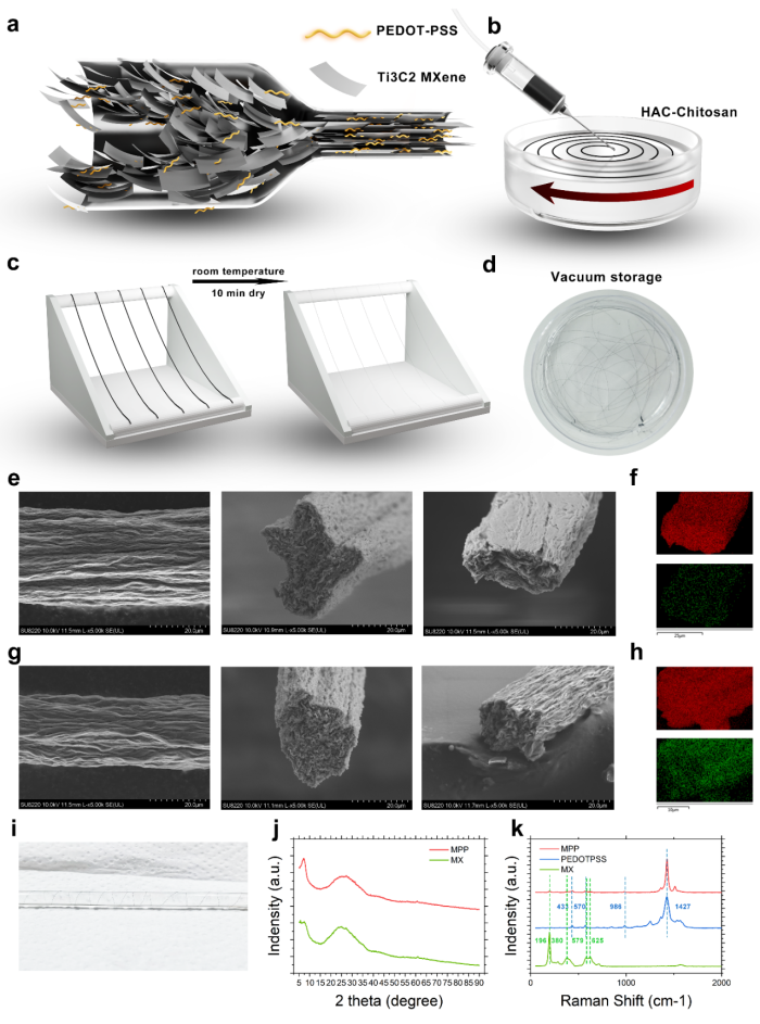
View the interface → Source figure · 2 views ?2R Complete Figure 1: fibre electrode manufacture, structural schematics and microscopy of MXene and MXene/PEDOT:PSS composite fibres.
SIAT liquid-crystal MXene/PEDOT:PSS fibre electrode Wet-spun fibre electrode in which MXene nanosheets are aligned in a nematic liquid-crystal state, with an MXene/PEDOT:PSS composite version. Du Zhanhong's lab at SIAT: 43.16 ohm mm2 at 1 kHz, 11.81 mC/cm2 charge injection, cortical recording in mouse and deep brain stimulation in rat.
Other Preclinical evidence
Photo credit Gou et al., Advanced Functional Materials (2025), Fig. 1, via SIAT · Gou et al., Advanced Functional Materials (2025), Fig. 1, via SIAT · Copyright retained by source; owner-requested noncommercial reference . Original source image preserved byte for byte; all embedded panels, annotations and scale bars retained.
View the interface → Photograph · 2 views ?2R Assembled experimental system, including the external PCB and a close-up of the electrode tips.
SIMIT mosquito-mouthpart bionic neural probe (128-channel flexible array) 128-channel, 2.5 micrometer polyimide array delivered through the dura by sharpened tungsten shuttles in microtubule tracks, with a tactile sensor array that warns of vessels. Built by Tao Hu and Wei Xiaoling's group at Shanghai Institute of Microsystem and Information Technology; mouse recordings to 16 weeks.
Intracortical Preclinical evidence
Photo credit Zhou et al., Microsystems & Nanoengineering (2023) · Microsystems & Nanoengineering · Figure 1e · CC BY 4.0 . Photographic panel extracted from the published source; resized and re-encoded. Original scale bars and annotations retained; no hardware retouching.
View the interface → Source figure · 3 views ?2R SINANO Figure 3 documents insertion and the implanted fibre-array configuration.
SINANO neuron-scale all-hydrogel fibre electrode array 16-channel array of PEDOT:PSS hydrogel fibres wet-spun and annealed under mechanical constraint to 0.94 to 20 micrometer diameter, the size of a neuron. Single-unit tracking for 6 months in mouse motor cortex, no clear gliosis at 16 weeks. Zhang Ting's group at Suzhou Institute of Nano-Tech and Nano-Bionics (Matter, 2026).
Intracortical Preclinical evidence
Photo credit Wang Mingxu, Li Lianhui, Zhang Ting group / SINANO, CAS (2026) · Wang Mingxu, Li Lianhui, Zhang Ting group / SINANO, CAS (2026) · Source copyright retained; site-owner-requested noncommercial scholarly reference . Original source image copied intact, byte for byte; labels, panels and scale bars retained.
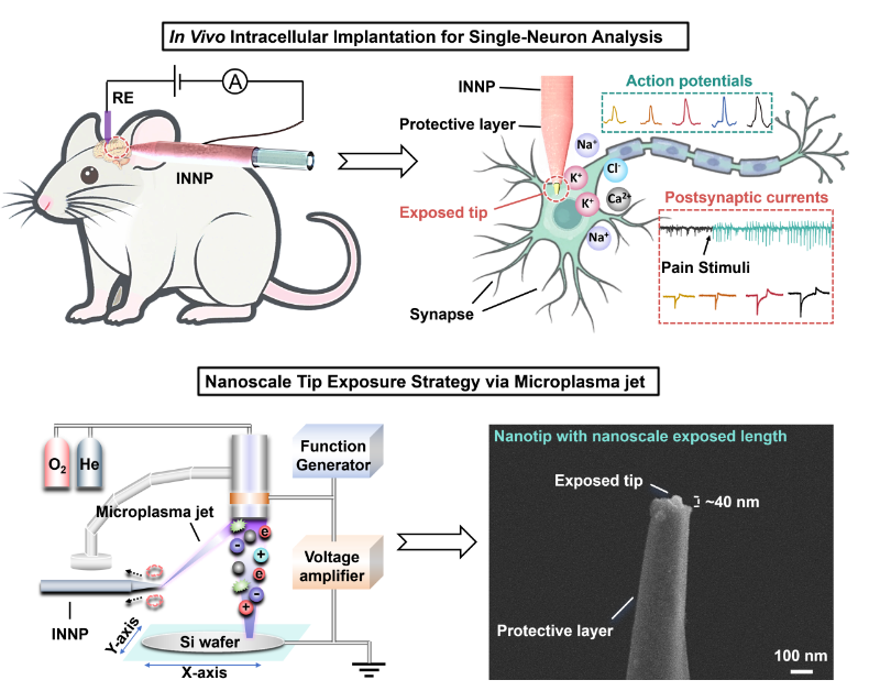
View the interface → Source figure ?2R SJTU’s IEEE MEMS 2026 INNP report includes the exposed tip SEM with the original 100 nm scale bar and approximately 40 nm exposed-tip annotation.
SJTU implantable nanoscale neural probe (INNP) for in vivo intracellular recording Probe with a tip about 100 nm across and a sensing region exposed over about 40 nm, so only the tip site sits inside one neuron. Records intracellular action potentials up to about 70 mV and synaptic currents in live mouse. Liu Jingquan's group at Shanghai Jiao Tong University (IEEE MEMS 2026 award paper).
Intracortical Preclinical evidence
Photo credit Du Zhiyuan and Liu Jingquan / Shanghai Jiao Tong University (2026) · Du Zhiyuan and Liu Jingquan / Shanghai Jiao Tong University (2026) · Source copyright retained; site-owner-requested noncommercial scholarly reference . Original source image copied intact, byte for byte; labels, panels and scale bars retained.
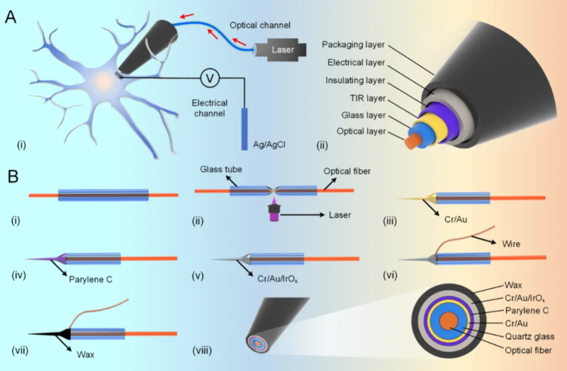
View the interface → Source figure · 2 views ?2R Primary SJTU release: probe layer architecture and fabrication, including the optical and electrical paths at the tip.
SJTU opto-electrophysiology nanotube probe with total-internal-reflection layer Laser-pulled nanotube probe with a nano optical fibre as the light channel inside and iridium oxide as the electrical channel outside, plus a total-internal-reflection layer that removes the photoelectric artifact. Intracellular recording and light stimulation in hippocampal neuron cell-line experiments. Liu Jingquan's group at Shanghai Jiao Tong University (ACS Nano 2024).
Other Preclinical evidence
Photo credit Liu Jingquan group / Shanghai Jiao Tong University (2024) · Liu Jingquan group / Shanghai Jiao Tong University (2024) · Source copyright retained; site-owner-requested noncommercial scholarly reference . Original source image copied intact, byte for byte; labels, panels and scale bars retained.
DEVICE IMAGE Photograph pending verification C3R
Image unavailable · device record retained StairMed WRS ultra-flexible wireless BCI StairMed's Wireless Recording System, a skull-mounted battery-free implant with ultra-flexible penetrating electrodes, in early human trials at Huashan Hospital in Shanghai. Implant counts and several specifications are company disclosures.
Intracortical Human evidence
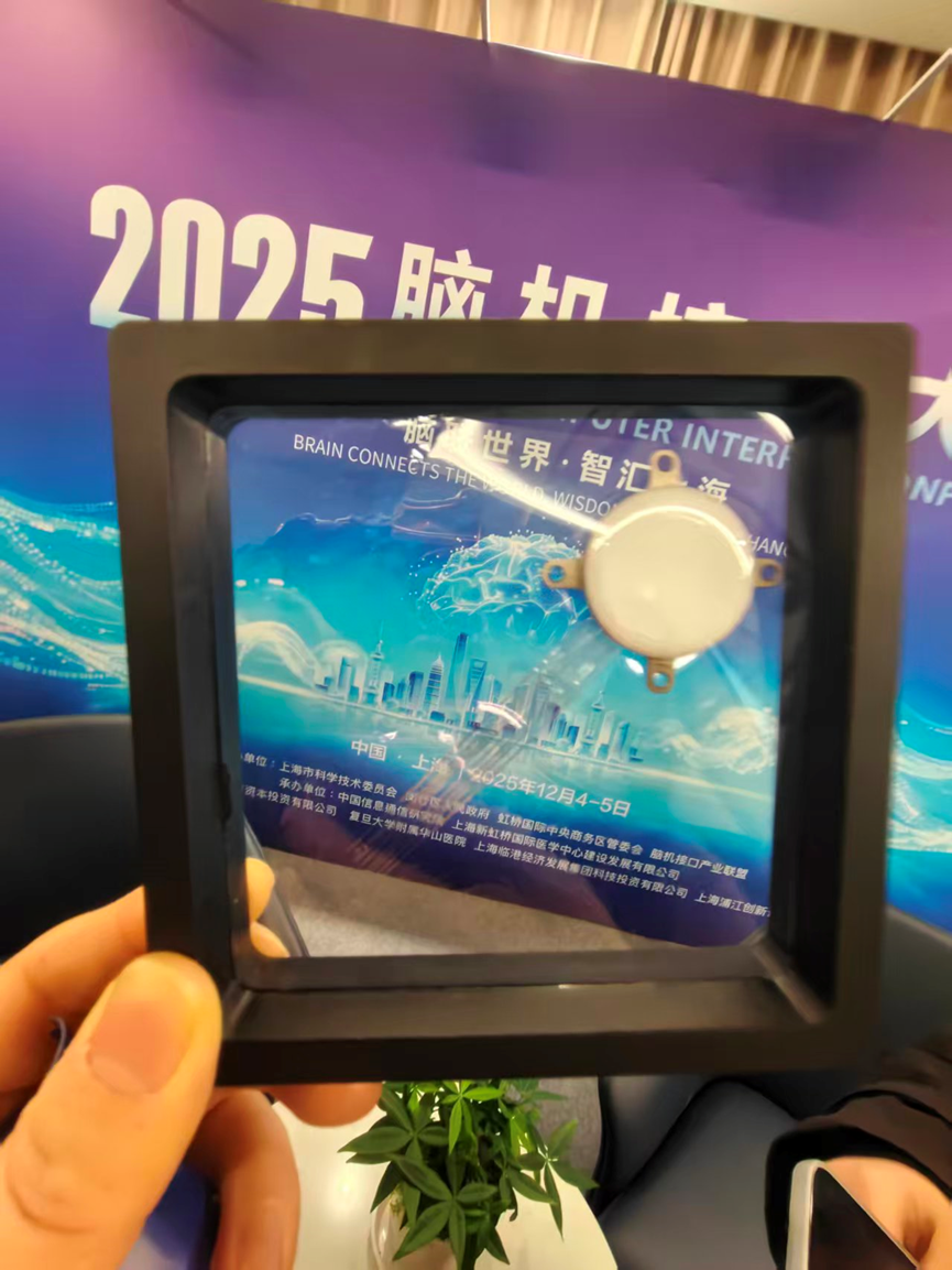
View the interface → Photograph C3R WRS02 display photographed at the 2025 launch, explicitly captioned as the second-generation 256-channel wireless implant system in the source.
StairMed WRS02 256-channel wireless BCI StairMed's second-generation 256-channel wireless BCI, launched in December 2025 and implanted from early 2026. Specifications are sparse and come from Chinese-language company disclosures and trade reporting.
Intracortical Human evidence
Photo credit StairMed product launch, via 科LAB report (2025) · StairMed product launch, via 科LAB report (2025) · Copyright retained by source; owner-requested noncommercial reference . Original source image preserved byte for byte; all embedded panels, annotations and scale bars retained.
View the interface → Electron micrograph L2R The tissue-facing microwire bundle of the Stanford microwire–CMOS interface. Scale bar: 500 µm.
Stanford microwire-CMOS bundles, 2020 A modular wire-to-chip interface with 135-251-wire mouse bundles, a 138-wire retina experiment and separate 8,640-wire connectivity demonstration. Chip pixels, readout capacity and implanted wires stay distinct.
Intracortical Preclinical evidence
Photo credit Obaid et al., Science Advances (2020), Figure 1C, CC BY-NC 4.0 · Original source · photographic panel · CC BY-NC 4.0 . Micrograph component selected from original PDF-embedded Figure 1C; 500 µm scale retained. Resized and re-encoded; no new image enhancement.
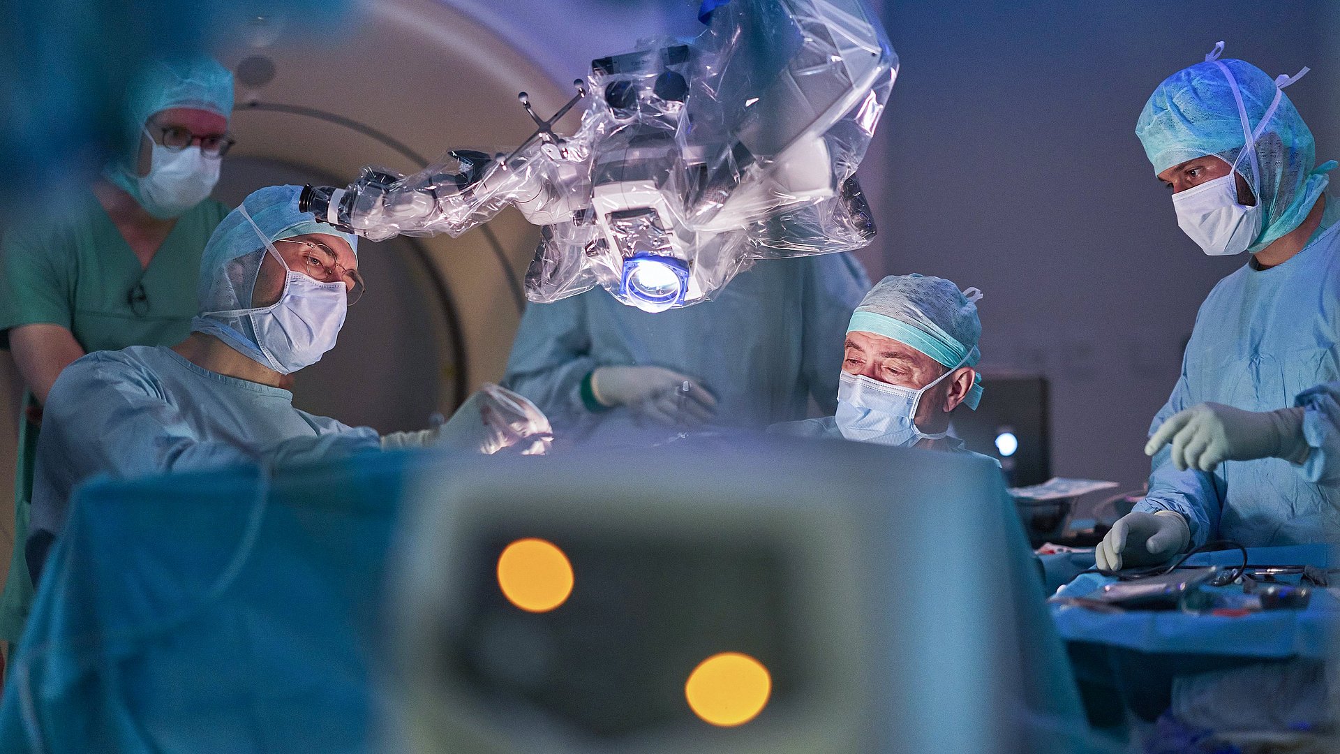
View the interface → Photograph ?3? TUM surgical team implants the four-array, 256-microelectrode BCI in the clinical study participant; photographed during the original 2025 research procedure.
TUM custom 256-microelectrode BCI implant Custom BCI with 256 microelectrodes implanted by a Technical University of Munich University Hospital team in a patient with quadriplegia, reported as Europe's first such procedure; aimed at smartphone and robotic-arm control.
Intracortical Human evidence
Photo credit Julia Bergmeister / TUM Klinikum · TUM University Hospital · Project press photograph · Project-specific editorial reuse with photographer credit . Original source photograph copied byte for byte to local storage; no crop, resizing or re-encoding. Source credit retained.
View the interface → Photograph ?2R The independent UFTE electrode fibers spread in distilled water. Scale bar: 1 mm.
Ultra-Flexible Tentacle Electrodes (UFTE) Swiss secondary coverage: 256 contacts on independent polyimide fibers in four bundles. Mechanical loop tethering separates shuttle removal from glue dissolution; the separate 512-channel logger stores data on SD rather than transmitting it.
Intracortical Preclinical evidence
Photo credit Yasar et al., Nature Communications (2024), Fig. 2a(v), CC BY 4.0 · Publisher · original figure · CC BY 4.0 . Select photo 2a(v) and retain 1 mm black scale bar.
View the interface → Photograph · 2 views C3R Manufacturer photograph of the Zhiran wireless implant platform.
Zhiran ultra-100-channel flexible BCI (LEAP trial) Beijing Zhiran's fully implanted wireless intracortical flexible BCI with over 100 channels, in a multicenter registration-style trial (LEAP) that launched at Tiantan Hospital in May 2026. Details come from Chinese hospital and state media releases and company statements.
Intracortical Human evidence
Photo credit Zhiran Medical official Chinese technology page · Zhiran Medical official Chinese technology page · Copyright retained by source; owner-requested noncommercial reference . Original source image preserved byte for byte; all embedded panels, annotations and scale bars retained.
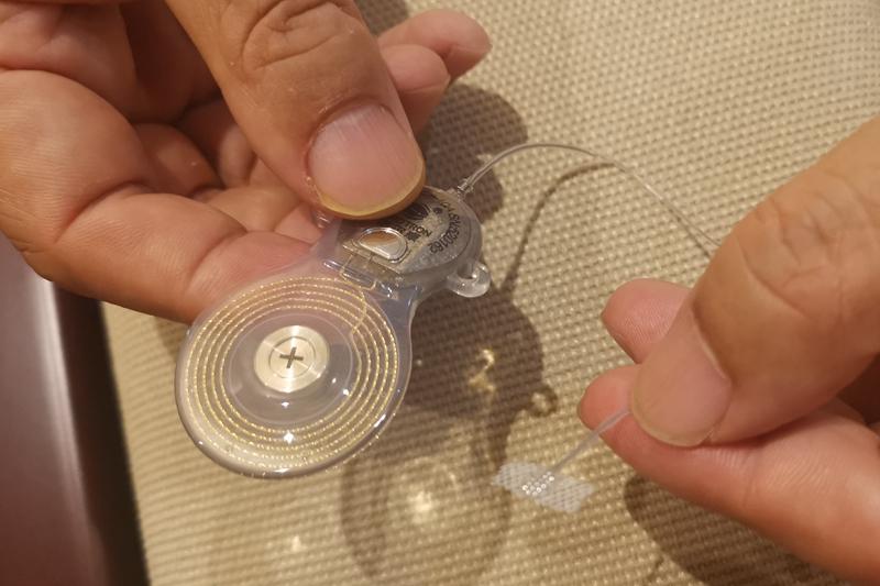
View the interface → Photograph L3? WIMAGINE implant viewed from its cortical-contact side, held between gloved fingers.
WIMAGINE (CEA-Clinatec) epidural wireless ECoG implant A fully implanted, wireless epidural ECoG system developed by CEA-Clinatec for human motor BCI research, emphasizing clinical robustness and long-term stability over single-unit precision.
Ecog Human evidence
Photo credit Sauter-Starace et al., Frontiers in Neuroscience (2019), Fig. 1A · Frontiers · Figure 1A · CC BY 4.0 . Photographic panel cropped from the original figure; resized and re-encoded. Original scale bars and annotations retained.
View the interface → Photograph C4K Precision Layer 7 array (right) beside a conventional ECoG strip (left).
Precision Layer 7 Cortical Interface Precision's thin-film cortical array. The 2025 paper reports a 1,024-channel version with 977 recording, 42 stimulation-optimized and five reference electrodes; FDA Layer 7-T clearance is separate from the future wireless BCI.
Ecog Human evidence
Photo credit Cummins et al., JNS Case Lessons (2026), Figure 2 · JNS Case Lessons · Figure 2 · CC BY-NC-ND 4.0 . Complete source image retained byte-for-byte, including every panel and annotation. No cropping, re-encoding, recoloring or synthetic enhancement.
View the interface → Source figure · 2 views C4A Original Tsinghua NEO rendering showing the epidural implant and external wireless unit.
NEO wireless epidural BCI (Neuracle, Tsinghua) A battery-free epidural ECoG implant for hand function in cervical spinal cord injury, developed at Tsinghua and commercialized by Neuracle. Reported as the first implantable BCI to receive market approval, in China in 2026. China entry, secondary region.
Ecog Human evidence
Photo credit Tsinghua University, original Chinese NEO announcement (2024) · Tsinghua University, original Chinese NEO announcement (2024) · Copyright retained by source; owner-requested noncommercial reference . Original source image preserved byte for byte; all embedded panels, annotations and scale bars retained.
View the interface → Photograph C3R INBRAIN graphene cortical array held with tweezers. Archival manufacturer photograph; source-image metadata dates to 2022, and the exact hardware revision is unspecified.
INBRAIN graphene cortical interface Thin-film graphene cortical surface interface from INBRAIN, used intraoperatively in a first-in-human study at Manchester. Company release; contact counts not published in the sources read.
Ecog Human evidence
Photo credit INBRAIN Neuroelectronics / Graphene Flagship, via EurekAlert! · Original source · physical interface · Public Domain — source-declared . Resized/re-encoded for web display only.
View the interface → Optical micrograph · 3 views L1R 240-channel NeuroGrid surface array; 1 mm scale retained.
NeuroGrid (PEDOT:PSS surface array) A 4 µm thick organic electrode array with 10 × 10 µm sites on 30 µm pitch that records single-neuron action potentials from the brain surface. NYU and collaborators, 2015.
3D model available Ecog Research evidence
Photo credit Khodagholy et al., Science Advances (2016), Figure 1 · Science Advances · Figure 1A · CC BY-NC 4.0 . Photographic panel extracted from the published source; resized and re-encoded. Original scale bars and annotations retained; no hardware retouching.
View the interface → Photograph · 3 views ?2R Original 14 × 14 micro-ECoG array photograph, preserving 500 µm overview and 150 µm inset bars.
Active flexible ECoG - Published hardware lineage
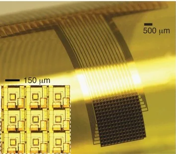
Channel Active Micro-ECoG A 14 x 14 multiplexed surface array with 196 platinum sites on 250 µm pitch and 29 interface wires on a roughly 25 µm film. Acute anesthetized rat auditory-cortex recordings, 2014.
The 2014 auditory-cortex configuration is the most recent documented design in this catalog, not a claim about present-day product availability.
3D model available Ecog Preclinical evidence
Photo credit Escabí et al., Journal of Neurophysiology (2014), Fig. 1B · Escabí et al., Journal of Neurophysiology (2014), Fig. 1B · Source copyright retained; owner-requested noncommercial reference . Source panel mechanically extracted; source annotations and scale bars retained. Resized and re-encoded.
Past versions and parallel branches (1) DEVICE IMAGE Photograph pending verification ?3?
Image unavailable · device record retained Dobelle cortical visual prosthesis A camera-driven visual cortex stimulator that gave blind volunteers phosphene images, built on a program that began in 1968. Reported by the Dobelle group in 2000.
Ecog Human evidence
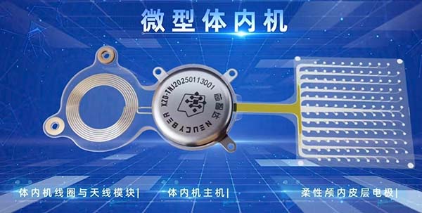
View the interface → Source figure · 2 views C3R Beinao-1 system image from the implanting hospital: coil, internal device and flexible cortical array.
Beinao-1 (CIBR, NeuCyber) A wireless, fully implanted, semi-invasive BCI with a 128-channel flexible electrode matrix, from the Chinese Institute for Brain Research, Beijing and NeuCyber NeuroTech. Entered registered clinical trials in 2026. China entry, secondary region.
Ecog Human evidence
Photo credit Peking University First Hospital, Beinao-1 clinical announcement (2025) · Peking University First Hospital, Beinao-1 clinical announcement (2025) · Copyright retained by source; owner-requested noncommercial reference . Original source image preserved byte for byte; all embedded panels, annotations and scale bars retained.
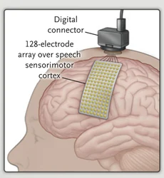
View the interface → Source figure · 2 views L3R 2021 source implant illustration: 128-contact PMT array and Blackrock percutaneous connector; schematic, not a hardware photograph.
High-density subdural ECoG array with percutaneous connector (PMT array, Blackrock connector) A subdural grid of flat disk electrodes made by PMT, wired through the skull to a Blackrock percutaneous connector. Two published speech implants used it: 128 electrodes at 4 mm pitch in 2021 and 253 electrodes at 3 mm pitch in 2023.
3D model available Ecog Human evidence
Photo credit Moses et al., NEJM (2021), Fig. 1 inset · Moses et al., NEJM (2021), Fig. 1 inset · Source copyright retained; owner-requested noncommercial reference . Source panel mechanically extracted; source annotations and scale bars retained. Resized and re-encoded.
View the interface → Photograph ?2R The real CLEAR graphene electrode array wrapped around a glass bar to demonstrate flexibility; 2014 device.
CLEAR transparent graphene micro-ECoG array A 16-site graphene surface array that combines electrical recording with light delivery and vascular imaging. Four graphene layers on parylene-C; rodent experiments, Wisconsin, 2014.
3D model available Ecog Preclinical evidence
Photo credit Park et al., Nature Communications 5, 5258 (2014), Figure 1c. DOI: 10.1038/ncomms6258 · Publisher · original figure · CC BY 4.0 . Photographic panel cropped from the original figure; resized and re-encoded. Original scale bars and annotations retained.
View the interface → Photograph · 3 views ?2R The 2024 UC San Diego transparent graphene array, including its flexible recording region and connector fanout.
High-density transparent graphene array (UC San Diego) Transparent graphene surface arrays up to 256 channels with 20 µm contacts and platinum nanoparticles. Combined electrical and calcium imaging in mouse visual cortex, 2024.
3D model available Ecog Preclinical evidence
Photo credit David Baillot / UC San Diego Jacobs School of Engineering · David Baillot / UC San Diego Jacobs School of Engineering · Institutional press image; attribution required; source copyright retained . Original source image copied intact, byte for byte; labels, panels and scale bars retained.
View the interface → Photograph C3R ABILITY neural-recording platform displayed on a transparent cranial model during its Wyss Center development program.
ABILITY Neurotech fully implantable communication BCI ABILITY Neurotech's fully implantable communication BCI: four cortical surface electrode arrays with wireless transmission for decoding into computer commands; in the recruiting INTRECOM clinical study at UMC Utrecht.
Ecog Human evidence
Photo credit © Wyss Center for Bio and Neuroengineering, ABILITY implantable neural recording system, via EurekAlert! (2022) · EurekAlert! · Wyss Center ABILITY image · CC BY-NC-ND 4.0 . Original source photograph copied byte for byte to local storage; no crop, resizing or re-encoding. Source credit retained.
View the interface → Photograph · 2 views L2R BISC chip held with tweezers, showing its thin, flexible form.
BISC wireless subdural CMOS array A 50 µm-thick wireless cortical chip with 65,536 physical electrodes and a selectable subset of up to 1,024 simultaneous channels. Published pig and non-human-primate recordings, December 2025.
3D model available Ecog Preclinical evidence
Photo credit Columbia Engineering; Jane Nisselson · Columbia Engineering; Jane Nisselson · Source copyright retained; site-owner-requested noncommercial scholarly reference . Original source image copied intact, byte for byte; labels, panels and scale bars retained.
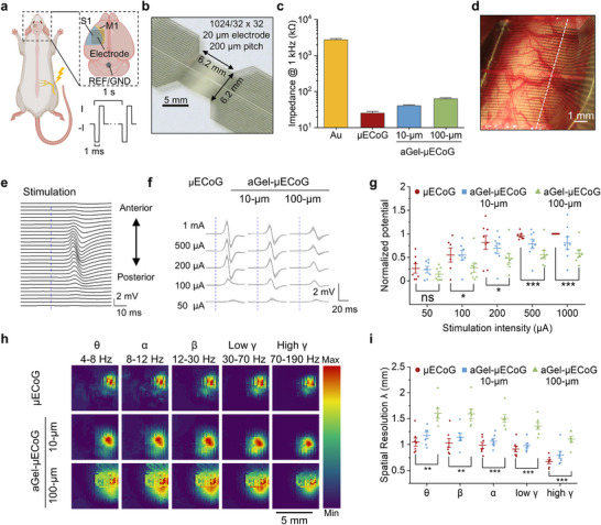
View the interface → Source figure ?2R Figure 3 of the paper: photograph of the 1024-channel array (b, 5 mm scale) and of the array on exposed rat cortex (d, 1 mm scale), with schematic and data panels a, c and e to i.
CIBR aGel-µECoG adhesive-hydrogel micro-ECoG array 6 micrometer polyimide micro-ECoG (64 or 1024 channels, 20 micrometer sites at 200 micrometer pitch) coated with a thin adhesive PVA/PTPM hydrogel that sticks to the brain without sutures and resists fibrosis. Wu Ting's lab at CIBR Beijing with Qiu Dong at ICCAS; 16 weeks subdural in rats (Advanced Science 2025).
3D model available Ecog Preclinical evidence
Photo credit Chinese Institute for Brain Research (CIBR) group and collaborators, Advanced Science (2025), Figure 3 · Advanced Science · complete Figure 3 (CC BY 4.0) · CC BY 4.0 . Complete source image retained byte-for-byte as served by PubMed Central (546 px wide rendition). No cropping, re-encoding, recoloring or synthetic enhancement.
View the interface → Source figure C3R Brain Interchange system and AirRay arrays documented in a 2022 canine-model study; the figure combines a system diagram with electrode photography.
CorTec Brain Interchange closed-loop BCI system CorTec's fully implantable, wireless, closed-loop neurotechnology platform: electronics between skull and scalp with silicone contact mats on the brain; first-in-human use in stroke rehabilitation reported from 2025.
Ecog Human evidence
Photo credit Schalk, Worrell, Mivalt, Belsten et al., Frontiers in Neuroscience (2022), Figure 1 · Frontiers in Neuroscience · Figure 1 · CC BY 4.0 . Original complete research figure copied byte for byte to local storage; no crop, resizing or re-encoding. Original panels, annotations and scale bars retained.
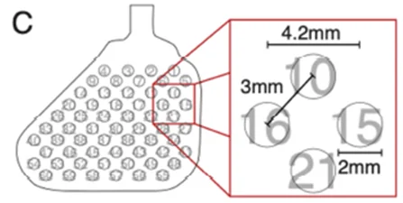
View the interface → Source figure · 2 views C3R Primary-study Figure 1C: Orion 60-electrode surface-array layout and enlarged contact arrangement. Schematic, with source dimensions retained.
Cortigent Orion visual cortical prosthesis Investigational 60-electrode cortical surface visual prosthesis from Cortigent, tested in a six-subject early feasibility study. Not FDA approved. Separate from the discontinued Argus II retinal implant.
Ecog Human evidence
Photo credit Oswalt et al. · Brain Stimulation 2021, Figure 1C · Oswalt et al. · Brain Stimulation 2021, Figure 1C · Source copyright retained; site-owner-requested research reference . Selected original source panel only; resized and re-encoded without upscaling. Original geometry and dimension labels retained.
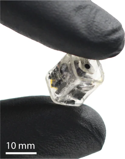
View the interface → Photograph L1R The packaged 2024 DOT epidural stimulator held between gloved fingers. Scale bar: 10 mm.
Pictured: DOT magnetoelectric epidural stimulator, 2024 . Other family versions differ.
DOT magnetoelectric platform - Research prototype and clinical program
Motif DOT magnetoelectric brain stimulator Pea-sized, battery-free wireless cortical stimulator from Motif Neurotech, shown acutely in a human in 2023 and now in an early feasibility study for treatment-resistant depression.
The photograph documents the 2024 research prototype. The Motif program is a separate clinical record; this image does not establish its later implant geometry.
Other Human evidence
Photo credit Woods, Singer et al., Science Advances (2024), Fig. 1A detail, CC BY 4.0 · Publication · original figure · CC BY 4.0 . Photo component selected from original PDF-embedded Figure 1A; original 10 mm scale retained. Resized and re-encoded; no hardware retouching.
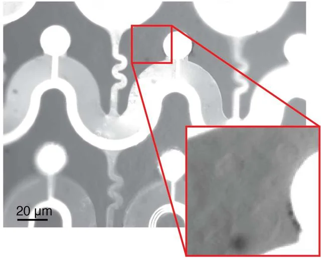
Related platform records (1) View the interface → Optical micrograph L2R The fenestrated tissue-facing Flex2Chip array with cerebellar cells in the same focal plane. Scale bar: 20 µm. The source inset has adjusted brightness and contrast.
Flex2Chip flexible-array CMOS interface Capillary-assembled suspended pads connect flexible neural arrays to a CMOS-MEA. Published 720/2,200-connection layouts, 1,024 simultaneous chip channels and acute 504-site ECoG use are kept distinct.
Ecog Preclinical evidence
Photo credit Zhao et al., Science Advances (2023), Fig. 4A, CC BY-NC 4.0 · Original source · physical interface · CC BY-NC 4.0 . Photographic panel cropped from the original figure; resized and re-encoded. Original annotations and scale bars retained where present.
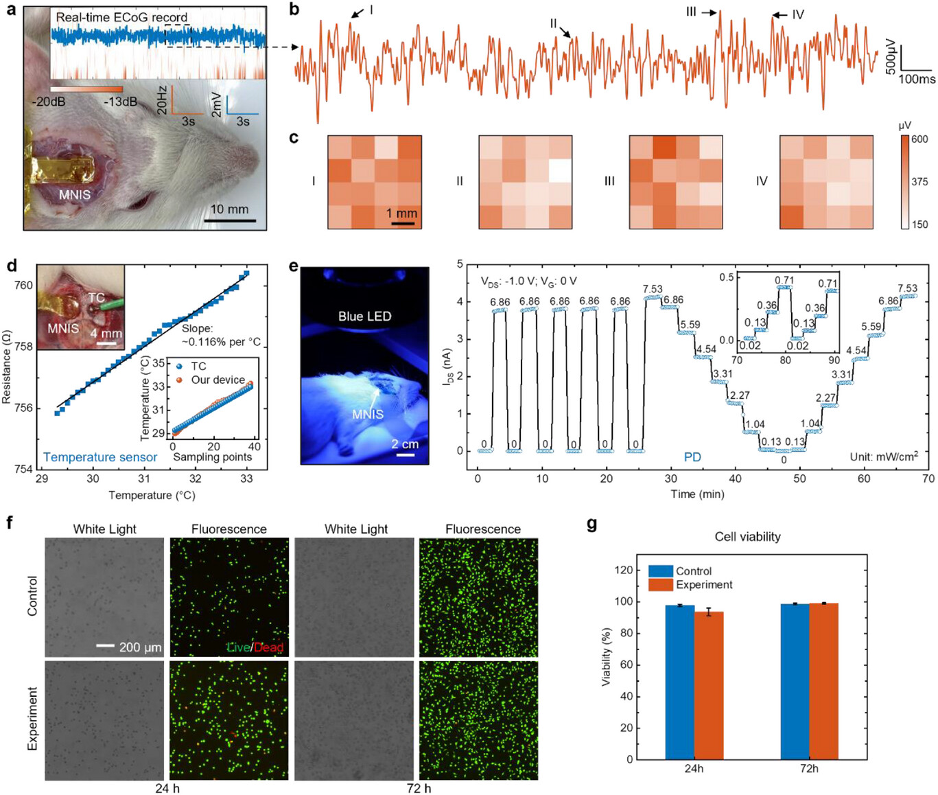
View the interface → Source figure L2R Figure 4 of the paper: photographs of the implanted device on a rat (a, 10 mm scale; d inset, 4 mm; e, 2 cm) alongside recording, temperature, photodetector and cell-viability data. Panels b, c, d, f and g are data plots and micrographs rather than device photographs.
Fudan MNIS active silicon-transistor multimodal cortical interface 4 x 4 array of silicon PMOS transistor nodes on a 31 micrometer flexible film that records ECoG by capacitive coupling, senses temperature with gold resistors and detects light with built-in photodiodes. Song Enming's group at Fudan University; acute rat recordings (Advanced Science 2025).
Ecog Preclinical evidence
Photo credit Song group and collaborators, Fudan University, Advanced Science (2026), Figure 4 · Advanced Science · complete Figure 4 (CC BY 4.0) · CC BY 4.0 . Complete source image retained byte-for-byte, including every panel and annotation. No cropping, re-encoding, recoloring or synthetic enhancement.
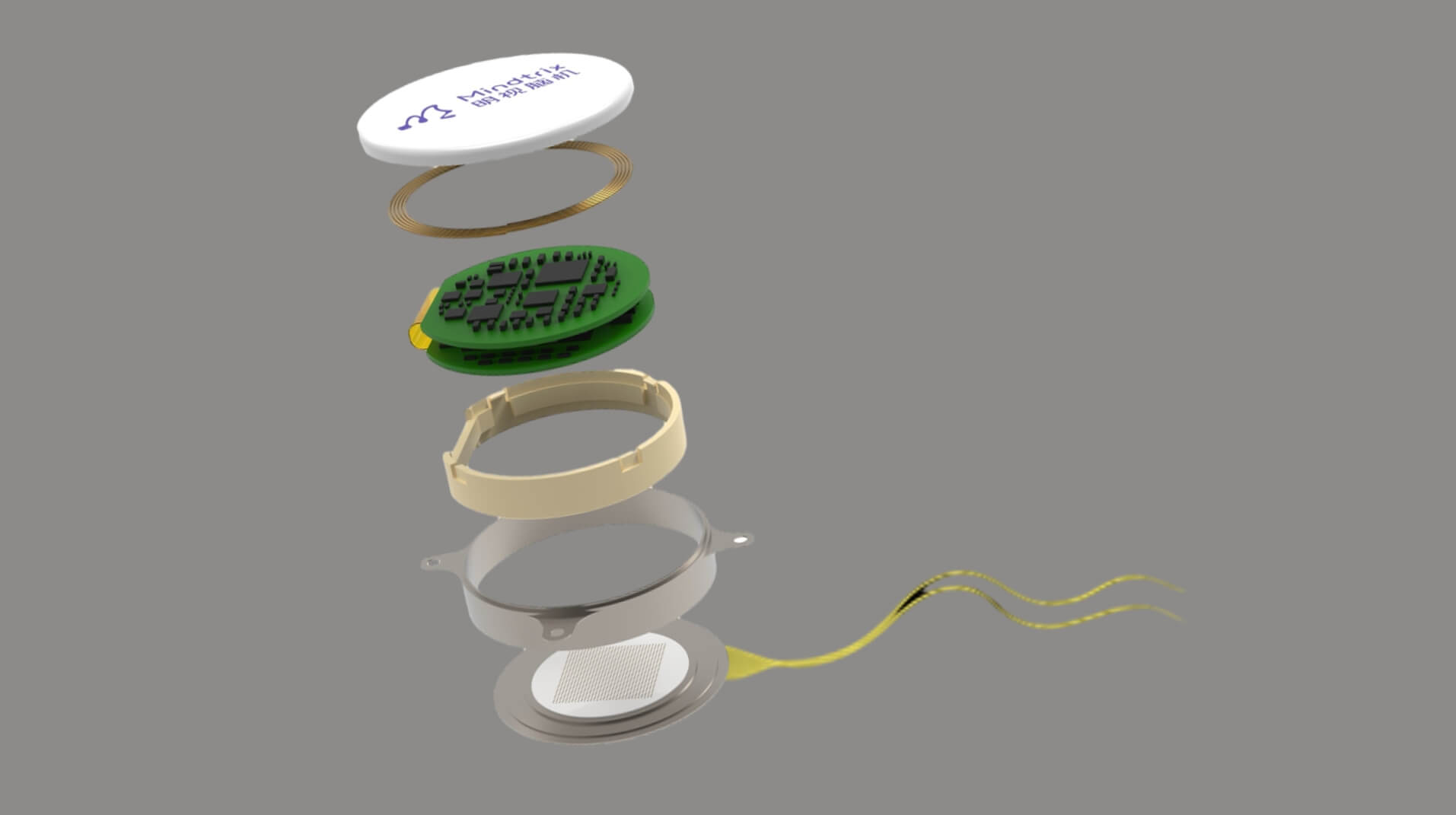
View the interface → Source figure · 2 views C3R Manufacturer architecture rendering of its fully implanted visual interface.
Mindtrix cortical visual reconstruction system Mindtrix's cortical stimulation system for visual reconstruction in blindness, with a first investigator-initiated human study reported in December 2025. Sources are Chinese-language press and the company site; study hardware is not fully described.
Ecog Human evidence
Photo credit Mindtrix official Chinese technology page · Mindtrix official Chinese technology page · Copyright retained by source; owner-requested noncommercial reference . Original source image preserved byte for byte; all embedded panels, annotations and scale bars retained.
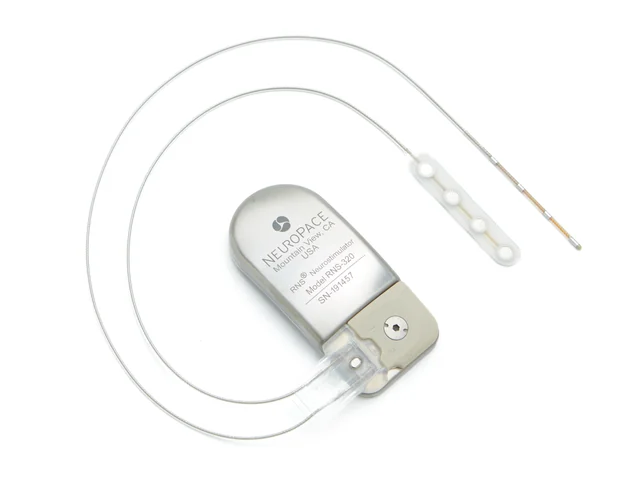
View the interface → Photograph · 2 views C4A NeuroPace RNS-320 neurostimulator with cortical strip and depth leads. Manufacturer press-kit photograph.
NeuroPace RNS System FDA-approved responsive neurostimulator that records ECoG through four-contact cortical strip and depth leads and delivers stimulation when programmed detections fire. Values come from the FDA summary and the published trials, scoped to the 2013 RNS-300M approval.
3D model available Ecog Human evidence
Photo credit NeuroPace · official press kit · NeuroPace · official press kit · Source copyright retained; site-owner-requested research reference . Original source re-encoded for display without upscaling; no hardware alteration.
View the interface → Source figure · 2 views C3R Complete Figure 1 of the 256-channel NeuroXess speech study: flexible cortical electrode placement, system architecture and signals.
NeuroXess 256-channel flexible ECoG BCI NeuroXess's 256-channel flexible cortical-surface array used for real-time motor and Mandarin speech decoding at Huashan Hospital. Reported in temporarily implanted epilepsy and tumor patients; some figures are company announcements.
Ecog Human evidence
Photo credit Qian et al., Science Advances (2025), Fig. 1 · Qian et al., Science Advances (2025), Fig. 1 · CC BY 4.0 . Original source image preserved byte for byte; all embedded panels, annotations and scale bars retained.
View the interface → Source figure · 2 views C3R Triple-F manufacturer rendering showing the cortical array, implant units, interconnecting lead and internal battery.
NeuroXess Triple-F fully implanted subdural BCI NeuroXess's fully implanted, fully wireless subdural BCI with a chest-pocket battery, in a registration trial at Huashan Hospital since July 2026. Most figures are company disclosures from Chinese-language sources.
Ecog Human evidence
Photo credit NeuroXess, Chinese GCP clinical-trial announcement (2026) · NeuroXess, Chinese GCP clinical-trial announcement (2026) · Copyright retained by source; owner-requested noncommercial reference . Original source image preserved byte for byte; all embedded panels, annotations and scale bars retained.
View the interface → Photograph L3R A flexible PtNRGrid cortical recording sheet held between fingers.
PtNRGrid platinum-nanorod surface arrays Reconfigurable cortical grids with 30 µm platinum-nanorod contacts in 6.6 µm parylene C. The 2022 paper reports 1,024- and 2,048-channel layouts with different pitch and coverage; a later IDE announcement is not commercial clearance.
3D model available Ecog Human evidence
Photo credit David Baillot / UC San Diego Jacobs School of Engineering · UC San Diego · official press photograph · Institutional press-image permission · credit required . Complete source image retained byte-for-byte, including every panel and annotation. No cropping, re-encoding, recoloring or synthetic enhancement.
View the interface → Optical micrograph · 2 views ?2R Physical contacts, traces and illuminated microLEDs of the small-format iEEG microdisplay.
PtNRGrid Recording and Display System PtNR surface electrodes laminated with GaN microLEDs to display cortical activity in the surgical field. Rat and pig proof of concept, including a 2,048-pixel display over 1,024 recording contacts and documented electrical interference.
Ecog Preclinical evidence
Photo credit University of California San Diego · UC San Diego · microdisplay close-up · Institutional press-image permission · credit required . Complete source image retained byte-for-byte, including every panel and annotation. No cropping, re-encoding, recoloring or synthetic enhancement.
View the interface → Source figure · 2 views L2R SCOPe’s primary packaging figure shows the real thin probe, enlarged chip/pixel photographs, and a labeled package cross-section.
SCOPe subdural CMOS optical probe Wired CMOS fluorescence imager and microLED stimulator with lensless reconstruction. Published abstract, supplement and 2023 preprint are distinguished; no wireless or single-cell claim.
Other Preclinical evidence
Photo credit Pollmann et al., Nature Electronics (2024), Figure 2 · Pollmann et al., Nature Electronics (2024), Figure 2 · Source copyright retained; educational reference reproduced at site owner request . Original source image preserved intact, byte for byte; all panels, scale bars and annotations retained.
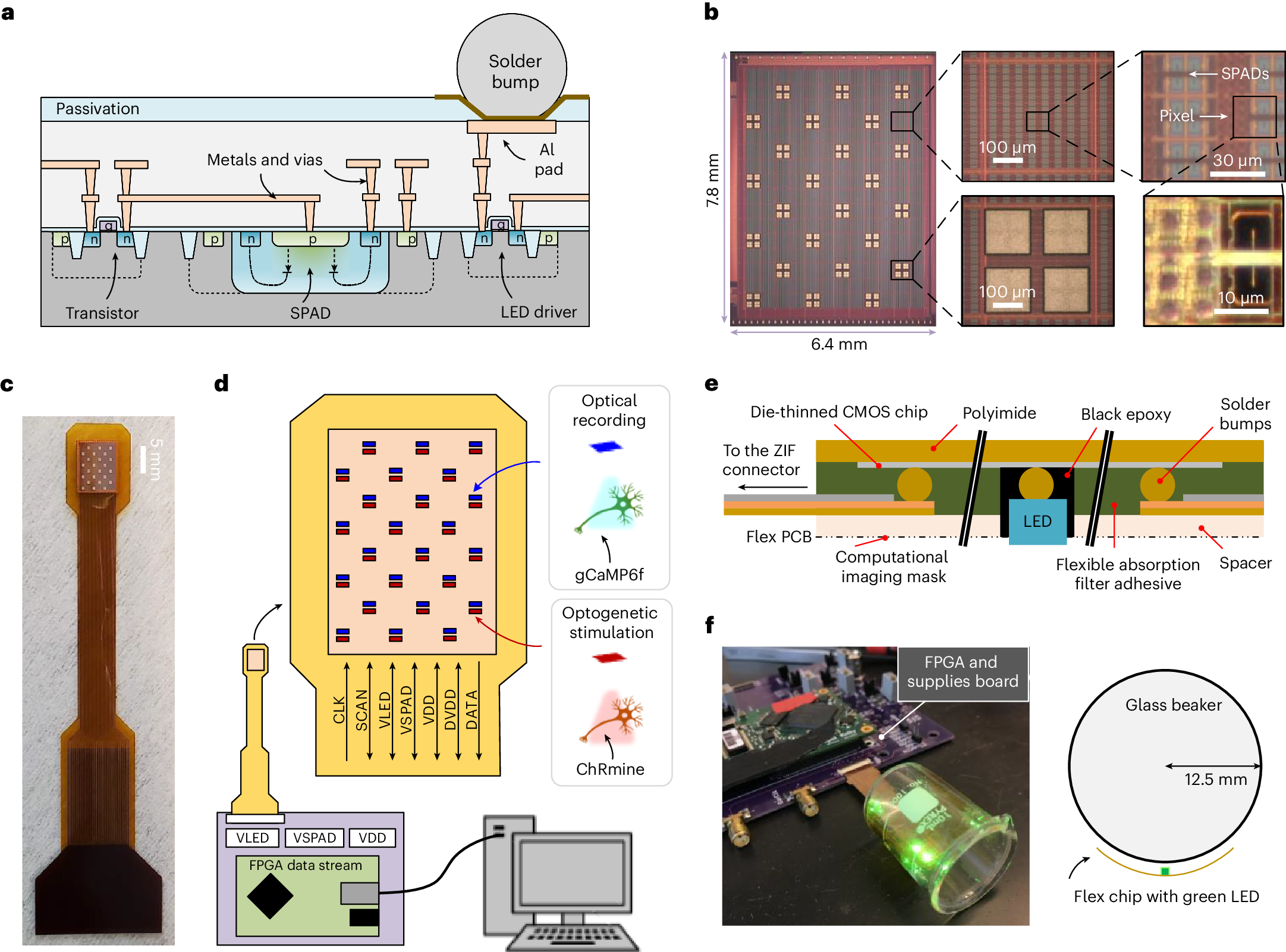
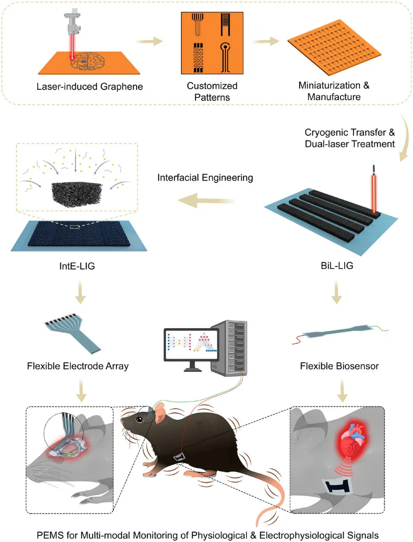
View the interface → Source figure · 2 views ?2R Original Figure 1 separates the wearable BiL-LIG sensor from the implanted IntE-LIG electrode; only the latter is the neural implant.
SIAT laser-induced graphene multimodal physiological-electrophysiological electrodes Two laser-induced graphene devices from Lu Yi's group at SIAT and SIBS: a stretchable BiL-LIG wearable sensor and an IntE-LIG neural electrode array with a conductive polymer-gold coating, 1 kHz impedance down 98.5%, 4 weeks of macaque and 12 weeks of mouse recording, with a mouse seizure closed loop.
Ecog Preclinical evidence
Photo credit Lu Yi group / SIAT and Shenzhen-Hong Kong Institute of Brain Science (2026) · Lu Yi group / SIAT and Shenzhen-Hong Kong Institute of Brain Science (2026) · Source copyright retained; site-owner-requested noncommercial scholarly reference . Original source image copied intact, byte for byte; labels, panels and scale bars retained.
View the interface → Photograph ?3? Integrated 256-channel µECoG implant with its surface array, reference lead and titanium electronics enclosure.
SIMIT 256-channel high-density micro-ECoG (64 electrodes/cm2) 16 x 16 gold-on-polyimide micro-ECoG array with 850 micrometer sites at 1250 micrometer pitch, in a titanium-housed implant with four Intan chips. 203 days in a Labrador, then intraoperative and short-term human motor-imagery decoding at Huashan Hospital. SIMIT with NeuroXess.
3D model available Ecog Human evidence
Photo credit Zhou et al., Advanced Science (2025), Figure 1b · Advanced Science · Figure 1b · CC BY 4.0 . Photographic panel extracted from the published source; resized and re-encoded. Original scale bars and annotations retained; no hardware retouching.
View the interface → Photograph · 2 views ?2R NeuroCam’s flexible cortical-surface electrode array, with its original 5 mm scale. The contact-bearing sheet is the neural interface; the curved support is a demonstration fixture.
Tsinghua NeuroCam 4096-channel TFT ECoG array Flexible, multiplexed cortical-surface array with 4096 metal-oxide thin-film-transistor channels at 44 sites per mm2, read through 128 lines. Sheng Xing's group at Tsinghua with Xuanwu Hospital; in vivo epilepsy-model recording.
Ecog Preclinical evidence
Photo credit Xing Sheng @ Tsinghua University, China; Science China Press · Tsinghua / Science China Press · NeuroCam photographs · Press image · use with credit . Optical photograph selected from panel (a) of the credited press image. Original scale bar retained; re-encoded without upscaling or hardware retouching. Press page permits use with credit; no Creative Commons grant is claimed.
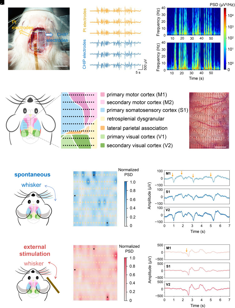
View the interface → Source figure ?2R Figure 4 of the paper: photographs of the CHIP-based array beside a commercial platinum array on rat cortex and of the array covering the cortex (scale bars as published), with recordings, spectrograms, a cortical-area schematic and activity maps in the remaining panels.
Tsinghua SIGS all-organic CHIP hydrogel ECoG array 128-channel ECoG array, 9 micrometers thick, made entirely of a conductive hydrogel (CHIP) printed on parylene, at 853 channels per cm2. Xu Xiaomin's group at Tsinghua Shenzhen International Graduate School with SIAT; rabbit recordings to 550 days (PNAS 2026).
Ecog Preclinical evidence
Photo credit Tsinghua Shenzhen International Graduate School group and collaborators, PNAS (2026), Figure 4 · PNAS · complete Figure 4 · CC BY-NC-ND 4.0 . Complete source image retained byte-for-byte, including every panel and annotation. No cropping, re-encoding, recoloring or synthetic enhancement.
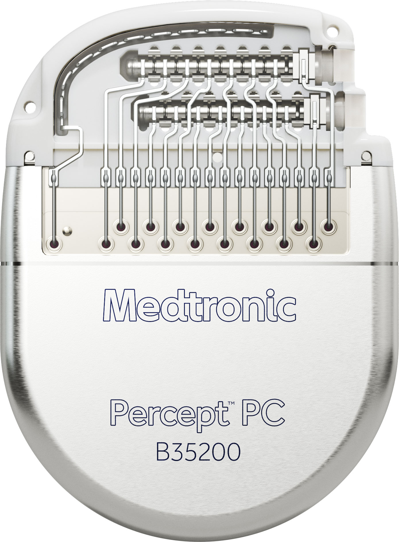
View the interface → Source figure · 3 views C4A Percept PC B35200 neurostimulator, intact Medtronic media-kit image.
Medtronic Percept and SenSight DBS FDA-approved deep brain stimulation family with sensing-enabled Percept neurostimulators and 8-contact SenSight directional leads. Values are scoped to the FDA record, Medtronic labeling and an independent technical report.
3D model available Dbs Human evidence
Photo credit Medtronic · Percept PC media kit · Medtronic · Percept PC media kit · Medtronic noncommercial educational website image license; no modifications . Source copied intact, byte for byte; no image alteration.
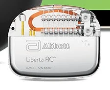
View the interface → Source figure · 2 views C4A Liberta RC model 62400 implant selected from Abbott's system image.
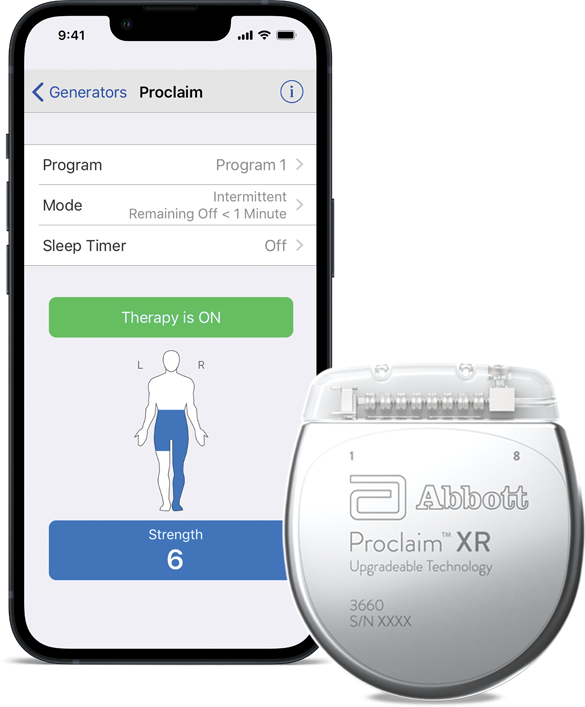
Abbott Liberta RC DBS system FDA-approved rechargeable DBS system from Abbott, approved by supplement S087 of PMA P140009 in January 2024. Many fields stay blank because the FDA record and company pages do not give them.
3D model available Dbs Human evidence
Photo credit Abbott · Liberta RC product page · Abbott · Liberta RC product page · Source copyright retained; site-owner-requested research reference . Selected original source panel only; resized and re-encoded without upscaling. Original geometry and dimension labels retained.
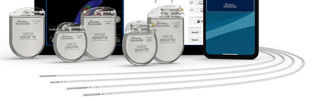
View the interface → Source figure · 4 views C4A Vercise Genus implantable pulse-generator family and contact-bearing DBS leads, selected from the current manufacturer system image.
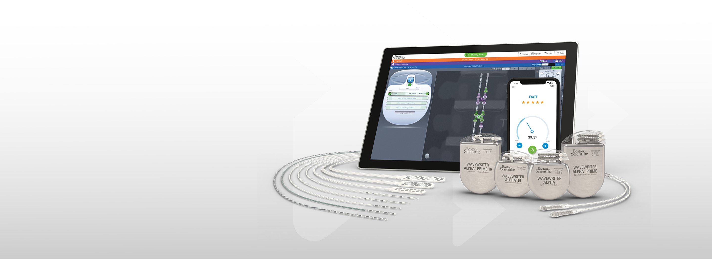
Boston Scientific Vercise Genus DBS FDA-approved Vercise Genus DBS family with 8-contact and 16-contact directional leads and Multiple Independent Current Control. Values rest on the FDA overview and company pages; the SSED was not read.
3D model available Dbs Human evidence
Photo credit Boston Scientific · Vercise Genus system · Boston Scientific · Vercise Genus system · Source copyright retained; site-owner-requested research reference . Implanted generator and lead region selected from the original manufacturer image; no geometry alteration or upscaling.
View the interface → Photograph L3? The long µSEEG electrode with its insertion stylet partly withdrawn, showing the thin flexible body. Scale bar: 1 cm.
µSEEG flexible stylet-guided depth electrode UC San Diego-led 2024 thin-film depth-electrode family: 32/64/128-contact variants, removable stylet, separate PEDOT human and PtNR primate configurations, and polymer/contact failure evidence.
Intracortical Human evidence
Photo credit Lee, Paulk et al., Nature Communications (2024), Fig. 1o, CC BY 4.0 · Publisher · original figure · CC BY 4.0 . Select source photo panel 1o only; preserve full lead, stylet and original scale.
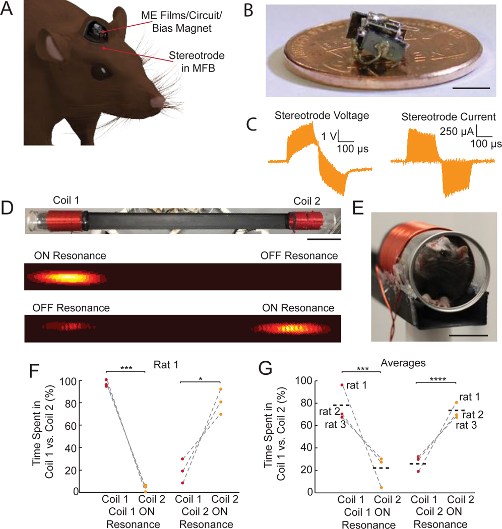
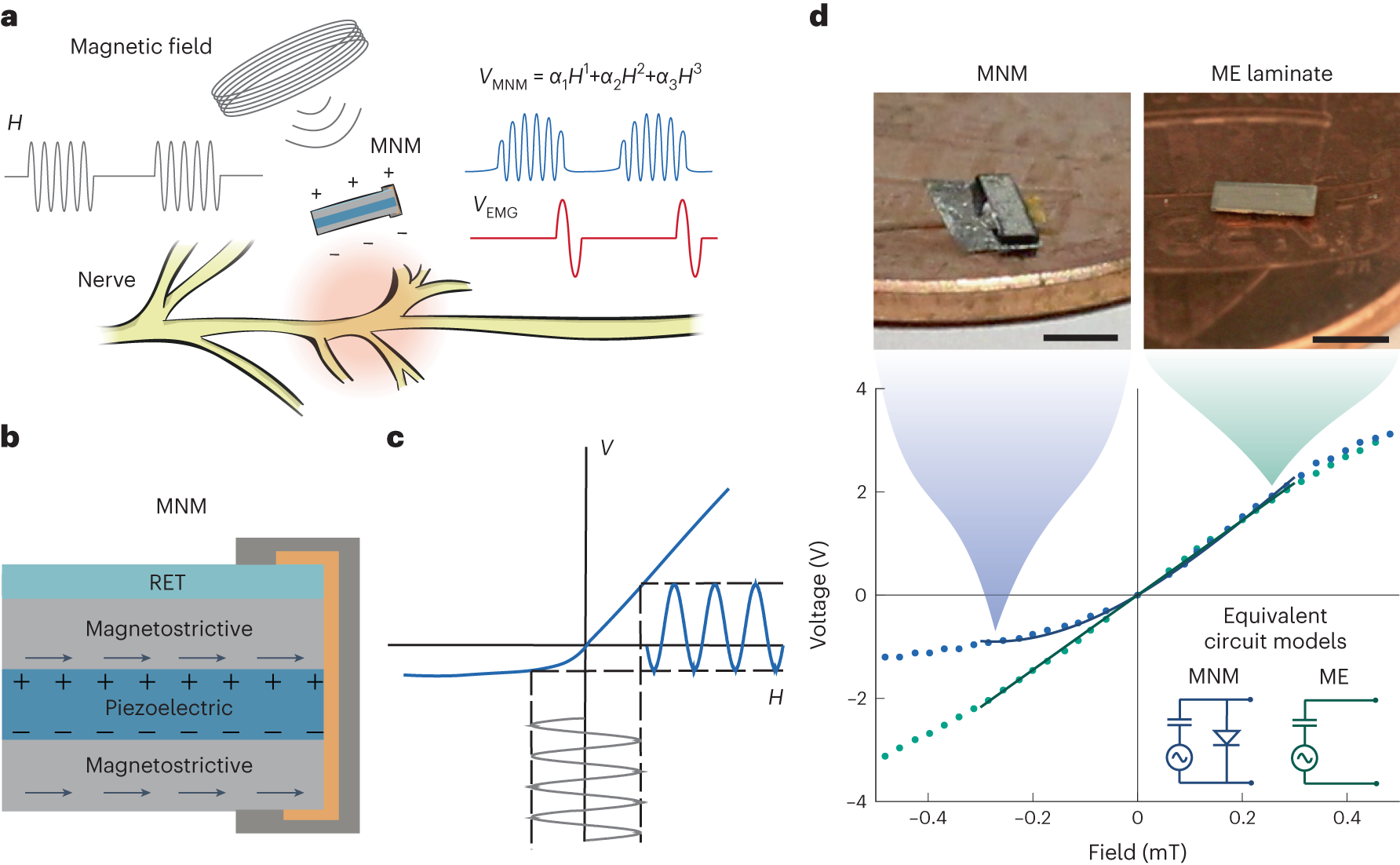
View the interface → Source figure L2R The original MFB place-preference experiment shows the compact two-film PZT circuit on a coin and its fully implanted placement linked to a stereotrode.
Two-film ME stimulators - Two configurations in one 2020 study
PZT Magnetoelectric Stimulator Two PZT/Metglas films, discrete rectifiers and a bias magnet in a 175-mm³ package drive rat MFB place preference. Acute behavioral evidence is separate from the PVDF Parkinson headstage.
The fully implanted PZT assembly is highlighted. The external PVDF headstage is a different configuration, not an earlier publication or an obsolete product.
Dbs Preclinical evidence
Photo credit Singer et al., Neuron (2020), MFB implant hardware figure · Singer et al., Neuron (2020), MFB implant hardware figure · Source copyright retained; educational reference reproduced at site owner request . Original source image preserved intact, byte for byte; all panels, scale bars and annotations retained.
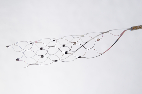
Past versions and parallel branches (1) View the interface → Photograph · 2 views C3R Stentrode scaffold and electrode contacts, with the recording lead extending to the right.
Stentrode (Synchron) A fully implanted endovascular BCI: a stent-electrode array in the superior sagittal sinus recording ECoG-like signals, trading spikes for catheter-based deployment.
3D model available Endovascular Human evidence
Photo credit Synchron Stentrode reference image supplied by the site owner; original photographer unspecified · Synchron · device background · User-supplied image; source copyright retained . Supplied image copied intact, byte for byte. All labels, panels, scale bars and embedded metadata retained.
View the interface → Photograph · 2 views C3R Wireless transmission device and subcutaneous placement from the 2024 endovascular sensor-removal study.
Nankai / Rongyuan interventional (stent-electrode) BCI Endovascular stent-electrode BCI from Duan Feng's group at Nankai University, with a subcutaneous wireless power and transmission unit. Sheep (2022), non-human primate (2023), sensor retrieval in sheep (2024) and a first human implant in a stroke patient (2025). Electrode geometry and materials are not published in the sources read.
Endovascular Human evidence
Photo credit Nankai University, original research announcement (2024) · Nankai University, original research announcement (2024) · Copyright retained by source; owner-requested noncommercial reference . Original source image preserved byte for byte; all embedded panels, annotations and scale bars retained.
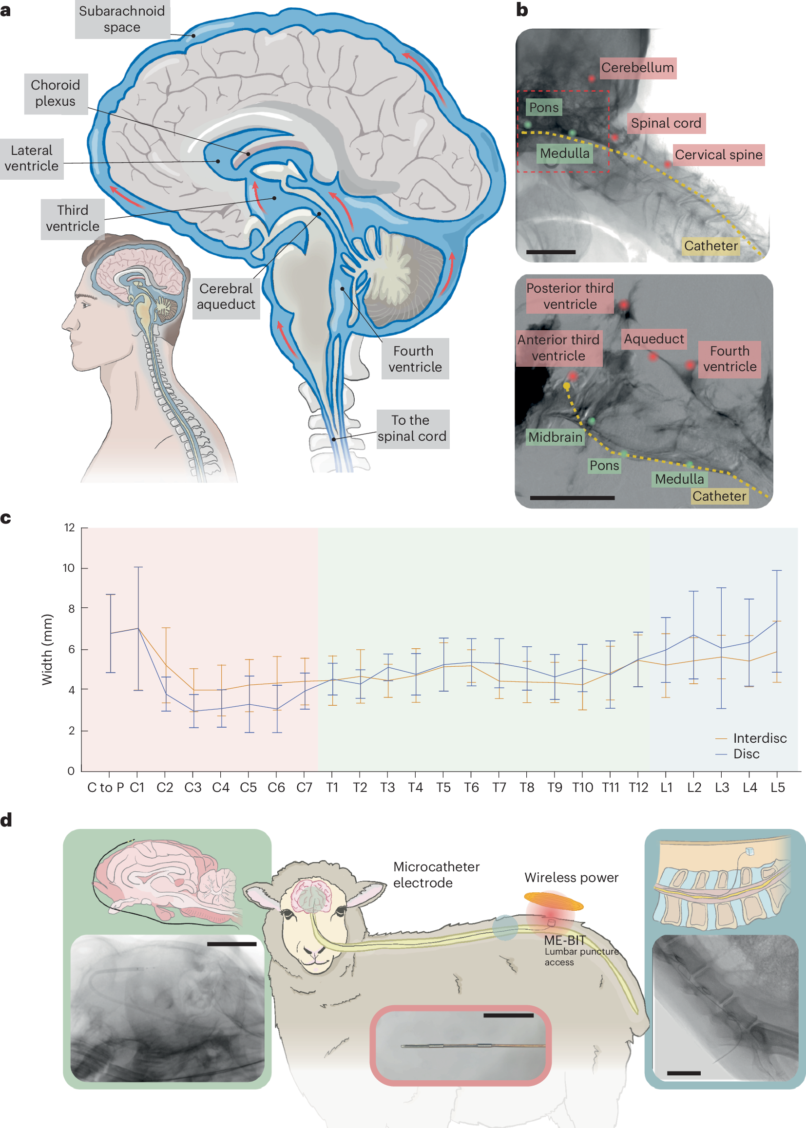
View the interface → Source figure · 2 views L2R The primary endocisternal study combines catheter placement images with a real electrode-tip photograph in the lower inset.
Endocisternal Magnetoelectric Interface Catheter electrodes navigated through cerebrospinal-fluid spaces, coupled to magnetoelectric bioelectronics. Full 2023 preprint specifications are separated from the 2024 published abstract and supplement.
Other Preclinical evidence
Photo credit Chen et al., Nature Biomedical Engineering (2024), Figure 1 · Chen et al., Nature Biomedical Engineering (2024), Figure 1 · Source copyright retained; educational reference reproduced at site owner request . Original source image preserved intact, byte for byte; all panels, scale bars and annotations retained.
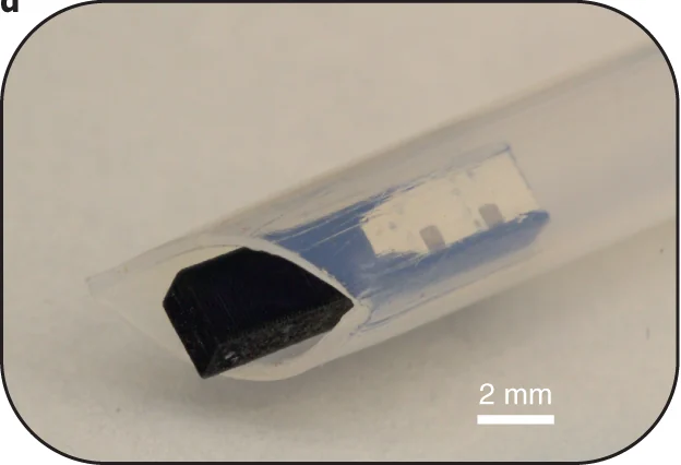
View the interface → Photograph L2R The encapsulated ME-BIT implant in its clear delivery sheath. Scale bar: 2 mm.
ME-BIT magnetoelectric nerve stimulator, 2022 2022 magnetoelectric-powered stimulation ASIC and packaged ME-BIT variants. Rodent direct contact, pig vascular leads and device delivery remain separate; chronic packaging, thrombosis and exposure-standard limits are explicit.
Endovascular Preclinical evidence
Photo credit Chen et al., Nature Biomedical Engineering (2022), Fig. 1d, CC BY 4.0 · Publisher · original figure · CC BY 4.0 . Select photo panel 1d only, retaining original rounded outline and 2 mm scale.
View the interface → Photograph · 2 views ?2R Unfolded intraventricular electrode, with conformal petals and surface contacts.
SIMIT silk-enabled intraventricular interface (IVI) Catheter-delivered 14 micrometer polyimide microelectrode array on a shape-memory silk fibroin scaffold that unfolds in cerebrospinal fluid and lies on the inner surface of the lateral ventricle. SIMIT and NeuroXess; recorded from the caudate head in Parkinsonian sheep for four weeks.
Other Preclinical evidence
Photo credit Liang et al., Nature Communications (2025), Figure 1 · Nature Communications · Figure 1e, lower photograph · CC BY 4.0 . Photographic panel extracted from the published source; resized and re-encoded. Original scale bars and annotations retained; no hardware retouching.
View the interface → Photograph ?3R CWRU/Ardiem standard self-sizing spiral cuff: nominal 4 mm diameter and four radial contacts. It wraps a peripheral nerve.
CWRU/Ardiem spiral cuff - Design and sensory-study records
CWRU/Ardiem self-sizing spiral nerve cuff A foundational, non-penetrating, self-sizing peripheral nerve cuff electrode design family (spiral/helical cuff) widely used for chronic stimulation and sometimes low-SNR recording.
These sheets cover the same self-sizing cuff design family. The standard four-contact photograph appears once; the sensory study is a deployment record, not a second cuff architecture. The ITIS 33-contact prototype has its own catalog entry.
Pni Human evidence
Photo credit Photo courtesy of Ardiem Medical, Inc.; Christie et al., Journal of NeuroEngineering and Rehabilitation (2017), Figure 1 · JNER · Figure 1 · CC BY 4.0 . Whole published photograph re-encoded without cropping or upscaling. Original Ardiem attribution retained.
Related study records (1) View the interface → Photograph · 3 views ?3R Fabricated 16-contact FINE in its open position, showing the tissue-facing contacts and reference strips. 10 mm scale retained.
FINE (Flat Interface Nerve Electrode) An extraneural cuff electrode that intentionally flattens/reshapes a peripheral nerve to bring fascicles closer to contacts, improving functional selectivity without penetrating the nerve.
3D model available Pni Human evidence
Photo credit Dweiri et al., Journal of Visualized Experiments (2016), Fig. 3A · Dweiri 2016 · Fig. 3A · Copyright reserved; educational reference use requested by site owner . Photographic panel mechanically cropped from the source; resized and encoded as WebP. Source annotations and scale bars retained; no generative changes.
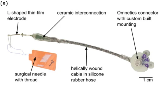
View the interface → Photograph ?3? TIME-4H assembly with thin-film electrode and helical lead; 1 cm scale retained.
TIME (Transverse Intrafascicular Multichannel Electrode) A flexible thin-film intrafascicular peripheral nerve interface inserted transversely through a nerve to access multiple fascicles, trading surgical complexity for selectivity.
3D model available Pni Human evidence
Photo credit Guiho et al., Sensors (2021), Fig. 2a · Sensors · Figure 2a · CC BY 4.0 . Photographic panel cropped from the original figure; resized and re-encoded. Original scale bars and annotations retained.
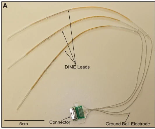
View the interface → Photograph · 3 views ?3? LIFE family example: Thota multi-LIFE DIME assembly with three leads, connector and ball ground. 5 cm source scale retained.
LIFE (Longitudinal Intrafascicular Electrode) A classic fine-wire intrafascicular peripheral nerve interface placed longitudinally within a fascicle for selective stimulation and compound action potential (CAP) recording.
3D model available Pni Human evidence
Photo credit Thota et al., Journal of Neuroscience Methods (2015), Fig. 3A; © 2014 Elsevier B.V. · Thota 2015 · Fig. 3A · Copyright © 2014 Elsevier B.V.; reserved-rights reference . Photographic panel mechanically cropped from the source; resized and encoded as WebP. Source annotations and scale bars retained; no generative changes.
View the interface → Photograph · 5 views ?3? ActiGait receiver, cable and nerve cuff, circa 2010 demonstration sample. Actual museum-object photograph; the sample is marked for demonstration use.
ActiGait implanted drop-foot stimulator (common peroneal nerve multi-contact cuff) A hybrid implanted-plus-external functional electrical stimulation (FES) system using a multi-contact cuff on the common peroneal nerve to restore ankle dorsiflexion during gait.
Pni Human evidence
Photo credit Science Museum Group Collection © The Board of Trustees of the Science Museum; Otto Bock Healthcare Products, object 2021-634 · Science Museum Group · ActiGait implant · CC BY-NC-SA 4.0 . Photograph resized and encoded as WebP; no retouching or generative changes.
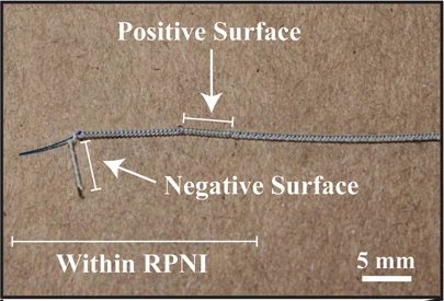
View the interface → Photograph · 3 views ?3? RPNI bipolar implanted electrode, contact close-up. Positive and negative exposed surfaces are labeled; 5 mm source scale retained.
Regenerative Peripheral Nerve Interface (RPNI) A muscle-mediated peripheral nerve interface (RPNI) used as a biological signal amplifier: reinnervated muscle grafts generate stable, high-SNR EMG for long-term prosthetic control without chronic intraneural electrodes.
Pni Human evidence
Photo credit Vu et al., Plastic and Reconstructive Surgery (2022), Fig. 2A inset; © American Society of Plastic Surgeons · Vu 2022 · Fig. 2A contact inset · Copyright © 2022 American Society of Plastic Surgeons; reserved-rights reference . Photographic panel mechanically cropped from the source; resized and encoded as WebP. Source annotations and scale bars retained; no generative changes.
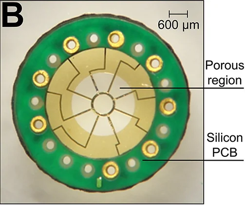
View the interface → Source figure · 2 views ?1R Redesigned mixed-porosity sieve, its assembly and microscopic contacts. Complete Figure 3.
Hyperflexible Regenerative Sieve A hyperflexible regenerative sieve electrode designed to promote neurovascular integration across a transected peripheral nerve interface (rat sciatic model).
Pni Research evidence
Photo credit Veith et al., Biomaterials (2021), Figure 3; © Elsevier Ltd. · Biomaterials · complete Figure 3 · © 2021 Elsevier Ltd. · source copyright retained . Complete PMC source figure copied intact, byte for byte. All panels, annotations and scale bars retained; no cropping, re-encoding, upscaling or synthetic enhancement.
View the interface → Optical micrograph ?1R Optical micrograph of the fabricated macro-sieve electrode and bonded micro-PCB; 2016 configuration.
Macro-Sieve Electrode A chronically implanted macro-sieve electrode with large transit zones enabling robust axon regeneration and selective stimulation in a rat sciatic nerve transection model.
Pni Research evidence
Photo credit MacEwan, Zellmer, Wheeler, Burton & Moran, Frontiers in Neuroscience (2016), Figure 1B. DOI: 10.3389/fnins.2016.00557 · Frontiers · original figure · CC BY 4.0 . Photographic panel cropped from the original figure; resized and re-encoded. Original scale bars and annotations retained.
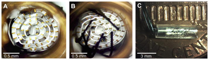
View the interface → Source figure · 2 views ?1R Microchannel interface assembly with integrated microwires, viewed from three sides. Complete Figure 9.
Regenerative Microchannel Interface A regenerative microchannel scaffold interface that guides axon regeneration into parallel channels and supports chronic recording/stimulation concepts (rat sciatic amputee model).
Pni Research evidence
Photo credit Srinivasan et al., Biomaterials (2015), Figure 9; © 2014 Elsevier Ltd. · Biomaterials · complete Figure 9 · © 2014 Elsevier Ltd. · source copyright retained . Complete PMC source figure copied intact, byte for byte. All panels, annotations and scale bars retained; no cropping, re-encoding, upscaling or synthetic enhancement.
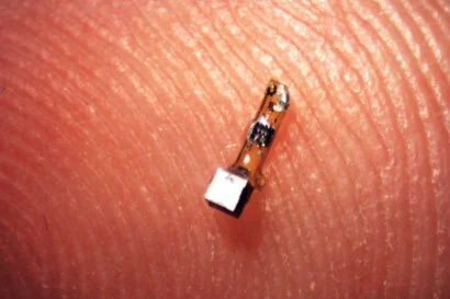
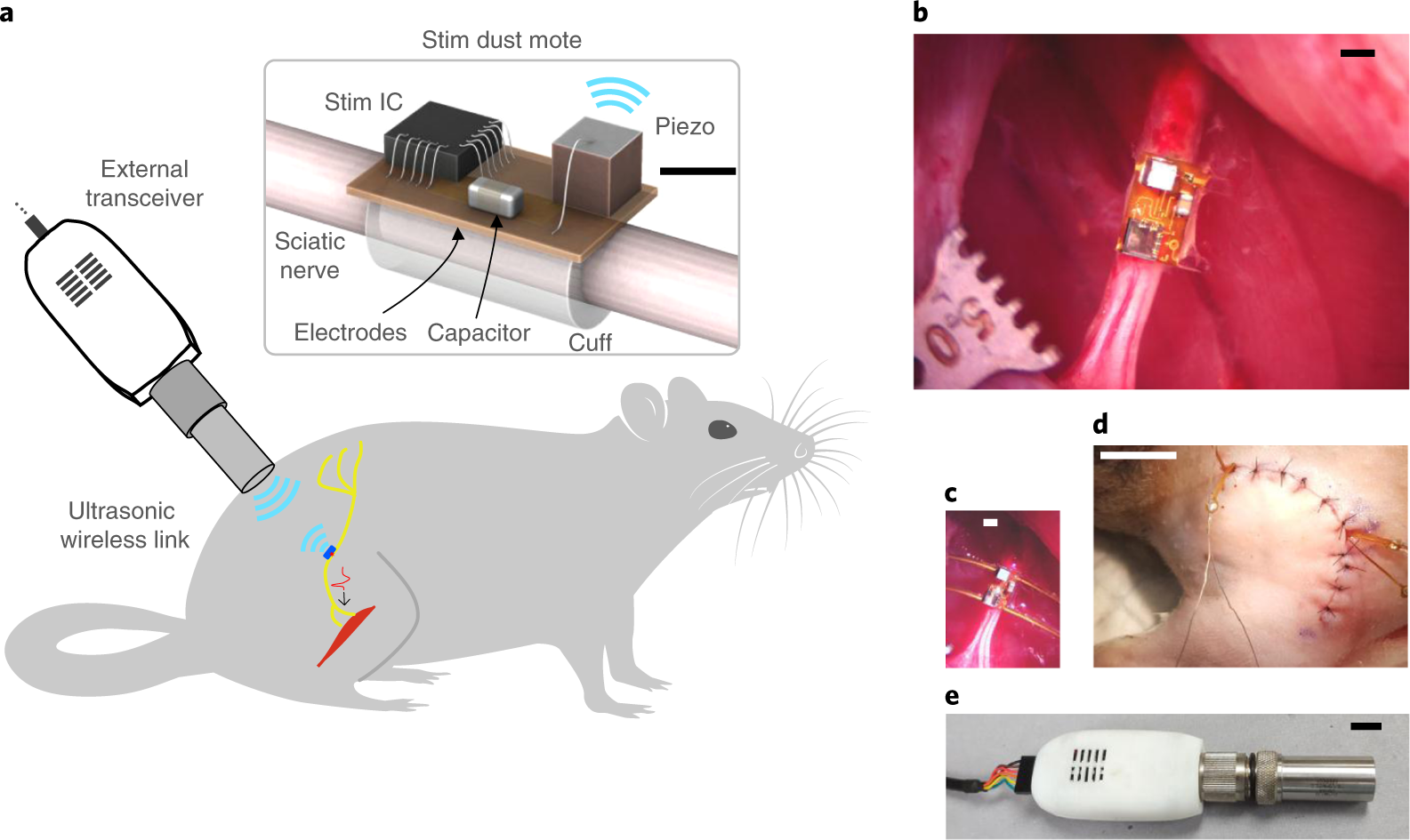
View the interface → Photograph · 3 views L2R Actual neural-dust mote with piezoelectric crystal and two-contact flexible circuit, on a fingertip.
Neural dust (ultrasonic backscatter mote) Ultrasonic recording mote: primary 2016 assemblies about 0.8 × 3 × 1 mm, one differential channel, 1.85-MHz interrogation and 10-kHz reconstructed signals. Rat nerve/muscle evidence; separate 1-mm-cube institutional claim.
Pni Preclinical evidence
Photo credit Ryan Neely / UC Berkeley (2016) · Ryan Neely / UC Berkeley (2016) · Source copyright retained; owner-requested noncommercial reference . Source figure resized and re-encoded; all panels, annotations and scale bars retained.
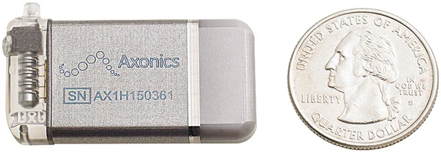
View the interface → Photograph C4A Axonics sacral neuromodulation pulse generator beside a U.S. quarter, from the 2020 ARTISAN-SNM study. Lead not shown.
Axonics sacral neuromodulation FDA-approved rechargeable sacral neuromodulation system with a four-contact tined lead. Values come from the 2019 FDA summary for the fecal incontinence indication.
Pni Human evidence
Photo credit Benson et al., Neurourology and Urodynamics (2020), Figure 1, CC BY-NC 4.0. · Original source · physical interface · CC BY-NC 4.0 . Full image retained; resized and re-encoded. Original annotations and scale bars retained where present.
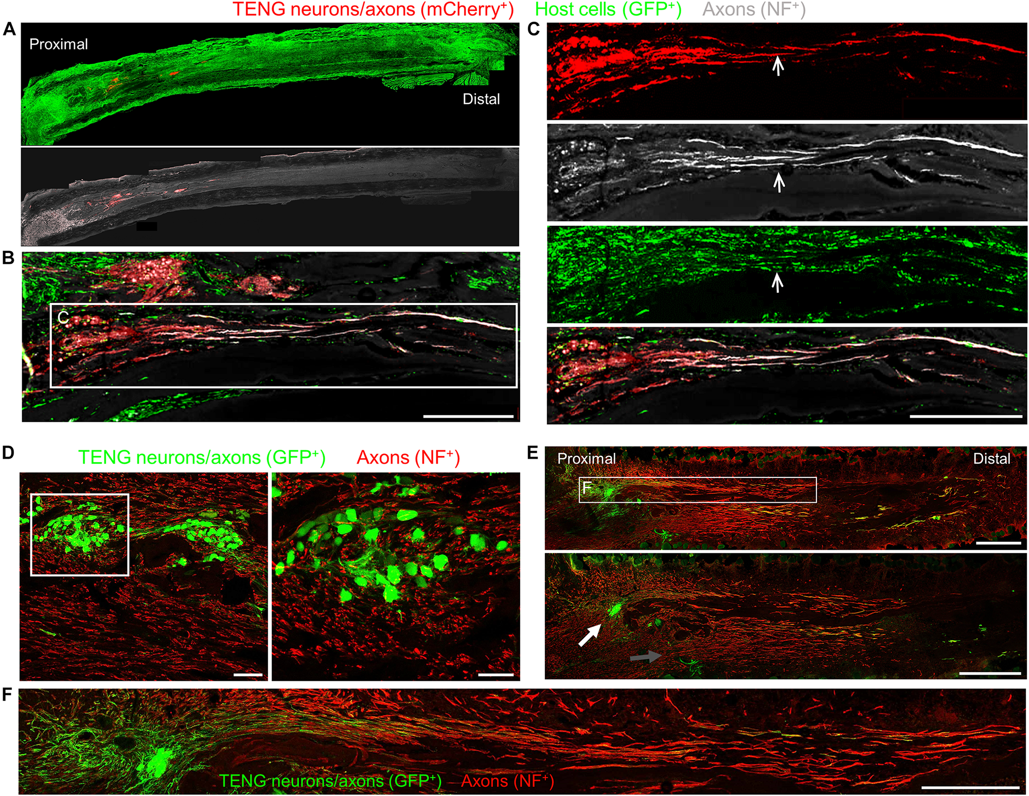
View the interface → Source figure ?2R Tissue-engineered nerve-graft axons and regenerating host nerve tissue visualized by fluorescence microscopy in preclinical experiments.
Axonova Medical tissue-engineered nerve graft Axonova Medical (UPenn spin-out): tissue engineered nerve grafts of living, aligned axon tracts spanning several centimeters, designed to guide long-distance axon regeneration after peripheral nerve injury.
Pni Preclinical evidence
Photo credit Katiyar, Struzyna, Morand, Burrell et al., Frontiers in Bioengineering and Biotechnology (2020), Figure 3 · Frontiers in Bioengineering and Biotechnology · Figure 3 · CC BY 4.0 . Original complete research figure copied byte for byte to local storage; no crop, resizing or re-encoding. Original panels, annotations and scale bars retained.
View the interface → Photograph · 2 views C4K The StimRouter implanted lead; the wearable transmitter is a separate component.
Bioness StimRouter PNS FDA-cleared peripheral nerve stimulation lead with an integrated receiver driven by an external transmitter, latest 510(k) read cleared February 2022. Thin: 510(k) text only.
Pni Human evidence
Photo credit Bioness / StimRouter · Bioness / StimRouter · Source copyright retained; site-owner-requested noncommercial scholarly reference . Original source image copied intact, byte for byte; labels, panels and scale bars retained.
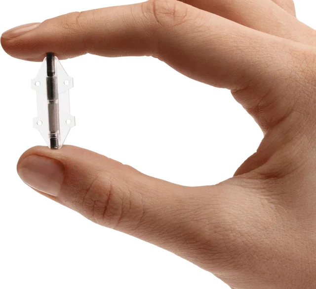
View the interface → Photograph · 2 views C4A BlueWind Revi implant held between fingertips, from the official manufacturer site.
BlueWind Revi tibial neuromodulation FDA-authorized wirelessly powered tibial nerve stimulator for urgency incontinence, from the De Novo review and 2024 510(k). Trial result values were not extracted.
Pni Human evidence
Photo credit BlueWind Medical · official implant photograph · BlueWind Medical · official implant photograph · Source copyright retained; site-owner-requested research reference . Selected original source panel only; resized and re-encoded without upscaling. Original geometry and dimension labels retained.
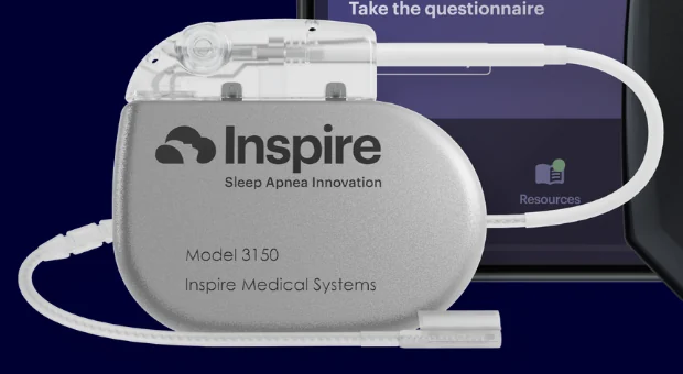
View the interface → Source figure · 2 views C4A Inspire V 3150 generator and its proximal lead loop, selected from the manufacturer's next-generation system image.
Inspire upper airway stimulation FDA-approved hypoglossal nerve stimulator for obstructive sleep apnea, newest version Inspire V approved August 2024. Thinly sourced from the FDA overview and supplement record; many cells stay blank.
Pni Human evidence
Photo credit Inspire Medical Systems · Inspire V source image · Inspire Medical Systems · Inspire V source image · Source copyright retained; site-owner-requested research reference . Selected original source panel only; resized and re-encoded without upscaling. Original geometry and dimension labels retained.
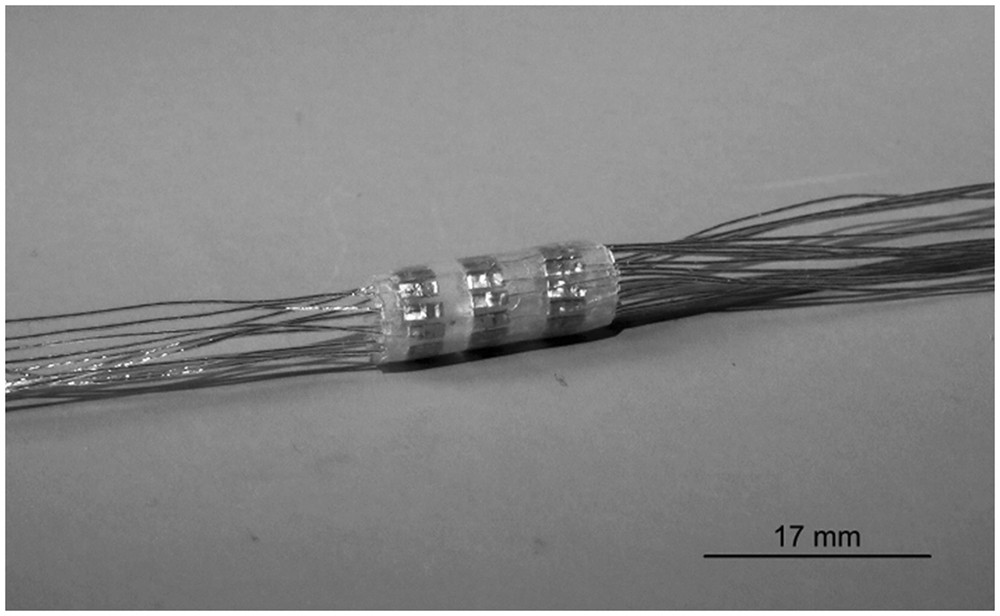
View the interface → Photograph ?1R ITIS/Ljubljana’s manufactured 33-contact spiral cuff, with contact rows visible through its sheath. Original 17 mm scale retained.
ITIS/Ljubljana 33-contact spiral cuff (2018 research prototype) A separate 33-contact platinum spiral-cuff prototype, photographed with its contact rows visible. Developed by the ITIS/Ljubljana team and tested on isolated porcine vagus nerve.
3D model available Pni Research evidence
Photo credit Rozman, Pečlin, Ribarič, Godec and Burja, Scientific Reports (2018), Figure 4 · Scientific Reports · manufactured cuff photograph · CC BY 4.0 . Complete published photograph copied intact, byte for byte, including its original scale bar.
View the interface → Source figure · 2 views C4A SenTiva model 1000 generator selected from the official system image.
LivaNova VNS Therapy SenTiva FDA-approved vagus nerve stimulation system for refractory epilepsy with the SenTiva generator that senses heart rate. Values come from the FDA supplement records, summary and LivaNova's spec sheet.
Pni Human evidence
Photo credit LivaNova · SenTiva manufacturer page · LivaNova · SenTiva manufacturer page · Source copyright retained; site-owner-requested research reference . Selected original source panel only; resized and re-encoded without upscaling. Original geometry and dimension labels retained.
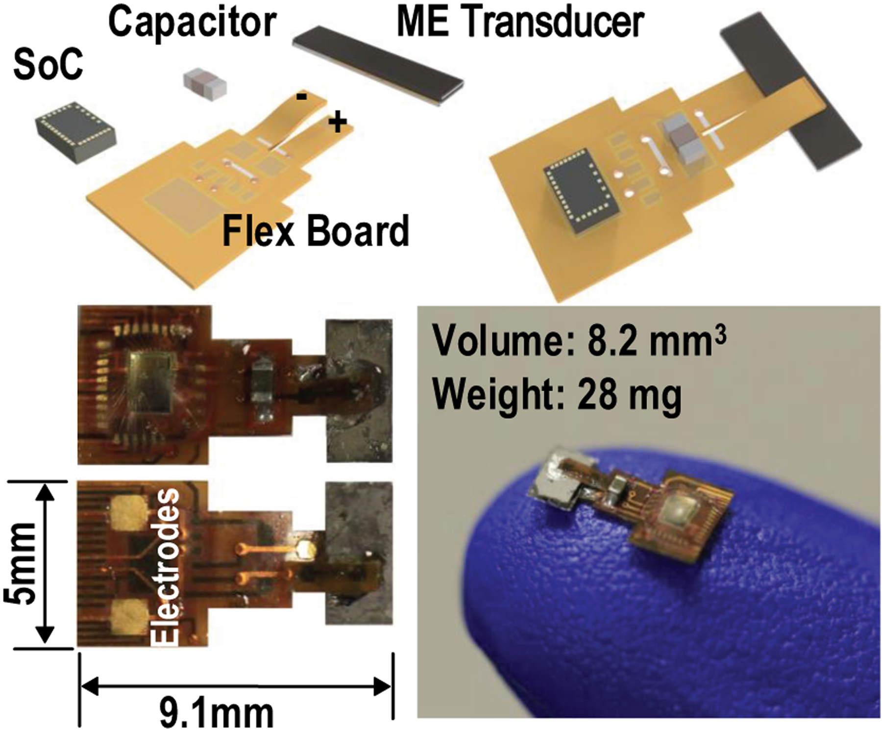
View the interface → Source figure · 2 views L2R MagNI’s primary construction figure shows the actual flex-board top and electrode underside, alongside the film/capacitor/SoC stack and fingertip photograph.
MagNI current-controlled ME implant, 2020 Published 8.2-mm³, 28-mg MagNI research stimulator: 250-kHz ME power, 1.5-mm² ASIC and programmable biphasic current. Hydra activation and a seven-day saline test are not spinal pain treatment.
Pni Preclinical evidence
Photo credit Yu et al., IEEE Transactions on Biomedical Circuits and Systems (2020), Figure 3 · Yu et al., IEEE Transactions on Biomedical Circuits and Systems (2020), Figure 3 · Source copyright retained; educational reference reproduced at site owner request . Original source image preserved intact, byte for byte; all panels, scale bars and annotations retained.
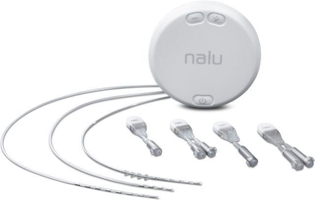
View the interface → Source figure C4K COMFORT study Figure 4: Nalu system with micro-IPG variants and four/eight-contact lead configurations.
Nalu neurostimulation system PNS FDA-cleared miniature peripheral nerve stimulator powered by a worn Therapy Disc, cleared 2019 with later 510(k)s through 2024. Thin: 510(k) text only.
Pni Human evidence
Photo credit Hatheway et al., Regional Anesthesia and Pain Medicine, Figure 4 · Hatheway et al., Regional Anesthesia and Pain Medicine, Figure 4 · CC BY-NC 4.0 . Original source image copied intact, byte for byte; labels, panels and scale bars retained.
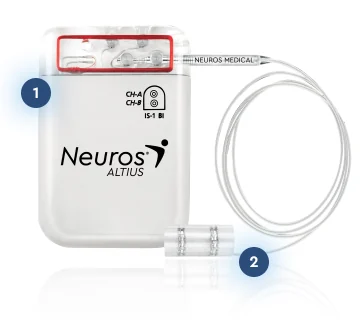
View the interface → Source figure · 2 views C4A Altius implanted pulse generator and nerve cuff, from the official manufacturer page.
Neuros Altius HFAC nerve block FDA-approved implanted nerve cuff system that blocks pain from an amputated limb with 5 or 10 kHz alternating current, approved August 2024. Values come from the FDA summary of safety and effectiveness.
Pni Human evidence
Photo credit Neuros Medical · Altius professionals · Neuros Medical · Altius professionals · Source copyright retained; site-owner-requested research reference . Original source re-encoded for display without upscaling; no hardware alteration.
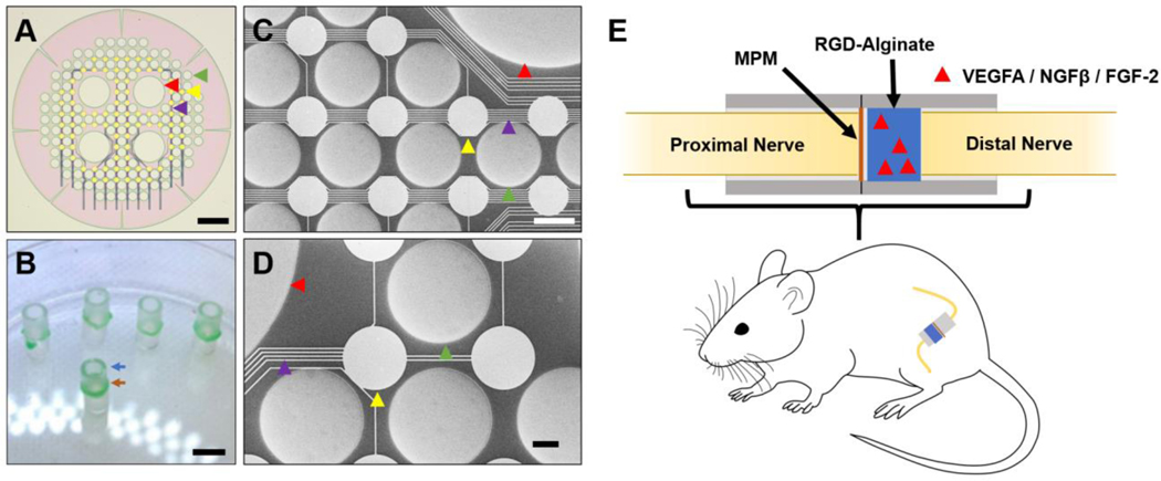
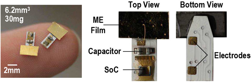
View the interface → Source figure L2R The primary platform overview shows packaged implants and their top and bottom surfaces, including the two tissue-facing electrode pads.
PUF-addressed ME multisite stimulator, 2021-2022 Rice-led 180-nm CMOS ME stimulation configuration with eight-bit PUF addresses and a shared transmitter. Bench and Hydra multisite demonstrations stay separate from rat nerve stimulation and proposed cardiac/spinal use.
Pni Preclinical evidence
Photo credit Yu et al., IEEE Journal of Solid-State Circuits (2022), Figure 2 · Yu et al., IEEE Journal of Solid-State Circuits (2022), Figure 2 · Source copyright retained; educational reference reproduced at site owner request . Original source image preserved intact, byte for byte; all panels, scale bars and annotations retained.
View the interface → Source figure L2R Panel D compares the actual self-rectifying magnetic neuromodulation metamaterial with a conventional ME laminate; panel B explains its engineered layer stack.
Self-Rectifying ME Implant Rice-led semiconductor-coated ME laminates for millisecond-timed peripheral stimulation. Schottky and p-Si/ZnO variants, low fabrication yield, short encapsulation life and external trigger hardware remain explicit.
Pni Preclinical evidence
Photo credit Chen et al., Nature Materials (2023), Figure 1 · Chen et al., Nature Materials (2023), Figure 1 · Source copyright retained; educational reference reproduced at site owner request . Original source image preserved intact, byte for byte; all panels, scale bars and annotations retained.
View the interface → Photograph C4A SetPoint's implanted pulse generator, shown beside a coin for scale. RESET-RA study device, 2024.
SetPoint vagus nerve stimulation for rheumatoid arthritis FDA-approved vagus nerve stimulator for rheumatoid arthritis, approved July 2025. Values come from the FDA summary of safety and effectiveness; trial efficacy numbers were not extracted.
Pni Human evidence
Photo credit Peterson et al., Bioelectronic Medicine (2024), Figure 2A, CC BY 4.0. Cropped from the original figure. · Publication · original figure · CC BY 4.0 . Photographic panel cropped from the original figure; resized and re-encoded. Original scale bars and annotations retained.
View the interface → Source figure ?2R CAS/SIAT design illustration of the ferroelectric bioelectronic vagus cuff and its self-rolling geometry.
SIAT ferroelectric bioelectronic interface (FBI) for vagus nerve Self-curling, suture-free hydrogel cuff for a 0.5 mm vagus nerve carrying a carbon nanotube and P(VDF-TrFE) ferroelectric strip that turns near-infrared light into nerve-like electrical pulses. Du Xuemin's group at SIAT; anti-inflammatory vagus modulation in rats (Advanced Materials, 2026).
Pni Preclinical evidence
Photo credit SIAT / Chinese Academy of Sciences, Du Xuemin group (2026) · SIAT / Chinese Academy of Sciences, Du Xuemin group (2026) · Source copyright retained; site-owner-requested noncommercial scholarly reference . Original source image copied intact, byte for byte; labels, panels and scale bars retained.
View the interface → Photograph · 2 views C4K MicroLead 2.0 held between fingers: original hardware photograph embedded in SPR's 2022 manufacturer sell sheet.
SPR SPRINT percutaneous PNS FDA-cleared temporary percutaneous peripheral nerve stimulation system with a wearable pulse generator, cleared January 2023 under 510(k) K223306. Thinly sourced; many cells stay blank.
Pni Human evidence
Photo credit SPR Therapeutics · MicroLead 2.0 sell sheet, page 1 · SPR Therapeutics · MicroLead 2.0 sell sheet, page 1 · Source copyright retained; site-owner-requested research reference . Original source re-encoded for display without upscaling; no hardware alteration.
View the interface → Source figure L2R The 2020 StimDust paper shows the real mote on the sciatic nerve in panel B, with the cuff and underside electrode arrangement illustrated in panel A.
StimDust ultrasonic nerve stimulator,2020 Published 1.7-mm³ battery-free stimulation mote, ultrasound-powered with backscatter status. Acute rat sciatic-nerve stimulation and 55-mm ex-vivo tissue link, not chronic clinical safety or neural recording.
Pni Preclinical evidence
Photo credit Piech et al., Nature Biomedical Engineering (2020), Figure 1 · Piech et al., Nature Biomedical Engineering (2020), Figure 1 · Source copyright retained; educational reference reproduced at site owner request . Original source image preserved intact, byte for byte; all panels, scale bars and annotations retained.
View the interface → Photograph · 2 views C3R ARC-IM implanted pulse generator photographed in the official ONWARD product page.
ONWARD ARC-IM implantable spinal stimulation Investigational implantable epidural spinal stimulation system for blood pressure instability after spinal cord injury, in the Empower BP pivotal study. Hardware specifications are not published in the sources read.
Scs Human evidence
Photo credit ONWARD Medical · ARC-IM manufacturer photograph · ONWARD Medical · ARC-IM manufacturer photograph · Source copyright retained; site-owner-requested research reference . Original source re-encoded for display without upscaling; no hardware alteration.
View the interface → Source figure · 2 views C4A Inceptiv neurostimulator front product image, retained intact from the Medtronic media kit.
Medtronic Inceptiv closed-loop SCS FDA-approved Medtronic spinal cord stimulator with closed-loop ECAP sensing, approved April 2024. Values rest on the FDA supplement record and Medtronic's announcement; the SSED was not read.
3D model available Scs Human evidence
Photo credit Medtronic · Inceptiv media kit · Medtronic · Inceptiv media kit · Medtronic media-kit copyright; source retained intact . Source copied intact, byte for byte; no image alteration.
View the interface → Source figure · 2 views C4A Evoke implant with two contact-bearing epidural leads, from Saluda's official product page.
Saluda Evoke closed-loop SCS FDA-approved closed-loop spinal cord stimulator that records ECAPs through two 12-contact leads, approved February 2022. Values come from the FDA summary of safety and effectiveness.
3D model available Scs Human evidence
Photo credit Saluda Medical · Evoke system · Saluda Medical · Evoke system · Source copyright retained; site-owner-requested research reference . Original source re-encoded for display without upscaling; no hardware alteration.
View the interface → Photograph · 4 views L2R Original EPFL e-dura photograph showing the transparent silicone carrier, soft contacts, interconnects and connector region.
e-dura (electronic dura mater) A soft implant with the shape and elasticity of the dura that carries electrodes and drug channels. EPFL, 2015: cortical recording in free-moving rats and supported stepping after paralysis with spinal electrical stimulation, drug delivery and rehabilitation.
Ecog Preclinical evidence
Photo credit EPFL / Alain Herzog © 2015 · EPFL / Alain Herzog © 2015 · Source copyright retained; owner-requested noncommercial reference . Source figure resized and re-encoded; all panels, annotations and scale bars retained.
View the interface → Source figure · 2 views C4A Proclaim XR generator and external patient-controller app; the phone is not implanted.
Abbott Proclaim XR spinal cord stimulation FDA-approved Abbott spinal cord stimulation family (Prodigy, Proclaim XR, Proclaim Plus, Externa), indications expanded to diabetic neuropathy and non-surgical back pain in 2023. Thin: hardware specs not in the FDA summary read.
Scs Human evidence
Photo credit Abbott · Abbott · Source copyright retained; site-owner-requested noncommercial scholarly reference . Original source image copied intact, byte for byte; labels, panels and scale bars retained.
View the interface → Source figure · 2 views C4A WaveWriter Alpha portfolio: implanted generators and electrode leads are shown alongside external controllers and adapters.
Boston Scientific WaveWriter spinal cord stimulation FDA-approved Boston Scientific spinal cord stimulation family (Spectra WaveWriter, WaveWriter Alpha, Alpha Prime), with the SOLIS trial for patients without prior back surgery. Lead contact counts and IPG dimensions are not in the FDA summary read.
3D model available Scs Human evidence
Photo credit Boston Scientific · Boston Scientific · Source copyright retained; site-owner-requested noncommercial scholarly reference . Original source image copied intact, byte for byte; labels, panels and scale bars retained.
View the interface → Photograph L2R Six magnetoelectric-powered spinal stimulators with their electrode leads. Authors' preprint prototype photograph, 2024. Scale bar: 1 cm.
Networked Magnetoelectric Spinal Stimulators 2025 network's spinal configuration: roughly 1-cm³ wireless battery-free IPGs, one electrode pair per node, six bench devices and up to four in acute pigs. LED/power/pacing demonstrations are not merged into that cohort.
Scs Preclinical evidence
Photo credit Woods, Alrashdan et al., Research Square preprint (2024), Fig. 3a, CC BY 4.0 · Original source · photographic panel · CC BY 4.0 . Photographic panel cropped from the original figure; resized and re-encoded. Original scale bars and annotations retained.
View the interface → Photograph · 3 views C4A Senza HFX iQ NIPG3000 implant photographed in the official product specification sheet.
Nevro Senza HFX iQ SCS FDA-approved 10 kHz spinal cord stimulation system with a 16-channel rechargeable IPG, approved October 2022. Values rest on the FDA supplement record and Nevro's prescriber information; the SSED and clinical data were not read.
3D model available Scs Human evidence
Photo credit Nevro · Senza HFX iQ IPG product specification sheet · Nevro · Senza HFX iQ IPG product specification sheet · Source copyright retained; site-owner-requested research reference . Original source re-encoded for display without upscaling; no hardware alteration.
View the interface → Source figure C3R Original Fudan system rendering from its 2025 industrial-exhibition announcement, showing the receiver, implant body, brain leads and spinal paddle.
Shenfu Jianxing brain-spine interface (Fudan) Fudan's brain-spine interface from Shenfu Jianxing: brain electrodes decode intent and drive spinal epidural stimulation in paraplegia. Four patients in a 2025 investigator-initiated study. Hardware specifications are not published; sources are Chinese-language releases and the ChiCTR registry.
Other Human evidence
Photo credit Fudan University, original Chinese CIIF research-exhibit release (2025) · Fudan University, original Chinese CIIF research-exhibit release (2025) · Copyright retained by source; owner-requested noncommercial reference . Original source image preserved byte for byte; all embedded panels, annotations and scale bars retained.
View the interface → Source figure C4A Argus II implant and retinal electrode array (A), with its external camera glasses and processing unit (B).
Argus II retinal prosthesis An epiretinal implant that sends camera-derived stimulation to a platinum electrode array tacked over the fovea. FDA humanitarian device approval in 2013; discontinued by its maker in 2019, leaving more than 350 users with an unsupported implant.
Retinal Human evidence
Photo credit da Cruz et al., Ophthalmology (2016), Figure 1 · Ophthalmology · complete Figure 1 · CC BY-NC-ND 4.0 . Complete source image retained byte-for-byte, including every panel and annotation. No cropping, re-encoding, recoloring or synthetic enhancement.
View the interface → Optical micrograph C4A 2 mm PRIMA photovoltaic retinal implant from the 2020 preclinical study. Dimension arrow retained.
Science PRIMA subretinal photovoltaic implant Subretinal 378-pixel photovoltaic implant with near-infrared camera glasses for geographic atrophy, with a 38-participant European study published in NEJM in October 2025.
Retinal Human evidence
Photo credit Muqit et al., PLOS ONE15(4):e0230713 (2020), Figure2 · Original source · physical interface · CC BY 4.0 . Right-hand photographic panel cropped; dimension marker preserved; resized/re-encoded for display.
View the interface → Photograph C4A Archival Nucleus specimen: Cochlear Freedom CI24RE receiver and electrode leads, photographed in 2009. Not the CI1000/Nexa generation.
Cochlear Nucleus implant system FDA-approved cochlear implant family, newest implant series CI1000 approved July 2025. Thinly sourced from the FDA supplement record and a Cochlear announcement; electrode specifications are not yet on the sheet.
Cochlear Human evidence
Photo credit Edwtie, Cochearimplants.JPG (2009), Wikimedia Commons, CC BY-SA 3.0. Cropped; the adapted photograph remains CC BY-SA 3.0. · Original source · photographic panel · CC BY-SA 3.0 . Photographic panel cropped from the original figure; resized and re-encoded. Original scale bars and annotations retained. The adapted photograph remains CC BY-SA 3.0.
View the interface → Source figure · 3 views C4A SYNCHRONY 2 cochlear implant product illustration from MED-EL, Innsbruck.
MED-EL cochlear implant system FDA-approved cochlear implant family with SYNCHRONY 2 as the current implant, indications expanded in October 2024. Values come from the FDA summary of safety and effectiveness; electrode specs are not on this sheet yet.
Cochlear Human evidence
Photo credit MED-EL · SYNCHRONY 2 manufacturer page · MED-EL · SYNCHRONY 2 manufacturer page · Source copyright retained; site-owner-requested research reference . Original source re-encoded for display without upscaling; no hardware alteration.
View the interface → Source figure · 2 views C4A HiRes Ultra 3D media image with the intracochlear electrode contacts visible along the distal lead.
Advanced Bionics HiRes Ultra 3D cochlear implant FDA-approved cochlear implant with a 16-contact HiFocus electrode, specified from the manufacturer's surgeon manual and the FDA PMA record P960058. Clinical data not extracted yet.
Cochlear Human evidence
Photo credit © Sonova AG / Advanced Bionics, HiRes-Ultra-3D-2 · © Sonova AG / Advanced Bionics, HiRes-Ultra-3D-2 · Source copyright retained; site-owner-requested noncommercial scholarly reference . Original source image copied intact, byte for byte; labels, panels and scale bars retained.
View the interface → Photograph · 2 views C3? Internal receiver-stimulator, lead and brainstem electrode array; source labels retained.
Auditory brainstem implant (ABI) A paddle electrode array placed on the cochlear nucleus of the brainstem to restore sound awareness when a cochlear implant cannot work. First implanted 1979, FDA approved in 2000 for NF2. The current US device has 21 platinum contacts on an 8.5 by 3.0 mm paddle.
Brainstem Human evidence
Photo credit Cochlear Americas © 2019, reproduced in Auditory Brainstem Implantation overview, Fig. 1B · Cochlear Americas © 2019, reproduced in Auditory Brainstem Implantation overview, Fig. 1B · Source copyright retained; owner-requested noncommercial reference . Source panel mechanically extracted; source annotations and scale bars retained. Resized and re-encoded.
View the interface → Photograph C3? Domestic auditory brainstem implant prototype, photographed during the Wu Hao team technology-transfer announcement.
Nurotron WH-01A auditory brainstem implant (Shanghai Ninth People's Hospital) Domestic Chinese auditory brainstem implant with 16 electrodes on a 5.5 x 3.5 mm silicone paddle, developed from Wu Hao's team at Shanghai Ninth People's Hospital. First implant January 26, 2022; NMPA innovative-device special review entry March 2024 (company report); NMPA approved the registration October 2026, a Chinese first for this device class.
Brainstem Human evidence
Photo credit Xi Rongpei / Shanghai Ninth People’s Hospital, via Shanghai Jiao Tong University (2021) · Xi Rongpei / Shanghai Ninth People’s Hospital, via Shanghai Jiao Tong University (2021) · Copyright retained by source; owner-requested noncommercial reference . Original source image preserved byte for byte; all embedded panels, annotations and scale bars retained.
View the interface → Electron micrograph · 3 views ?2R Empty microwell scaffold, sectioned and imaged at 45° tilt. 10 µm scale bar retained.
Science biohybrid cortical engraftment implant (microwell / waffle device) A surface cortical biohybrid implant: optogenetically enabled neurons seeded in a microwell scaffold on cortex, with neurites integrating into the brain to transmit information via optical stimulation.
Ecog Preclinical evidence
Photo credit Brown et al., Science Corporation (2024 preprint), Fig. 1b · Brown 2024 · Fig. 1b · Copyright reserved; educational reference use requested by site owner . Source figure image and its PDF soft mask extracted from manuscript page 16, composited on white, panel mechanically cropped; scale retained. No generative changes.
View the interface → Source figure · 2 views ?0R Science electronic biohybrid architecture concept: device-associated neurons and their brain connections. Company illustration, without a measured form factor.
Science neuronal-embedded electronic biohybrid interface (architecture concept) A company-described biohybrid architecture with neurons embedded in electronics; hardware geometry and complete-system performance remain not reported.
Other Theoretical evidence
Photo credit Science Corporation, Biohybrid neural interfaces (23 November 2024) · Science Corporation · architecture illustration · Copyright © Science Corporation; reserved-rights reference . Company source illustration retained intact; no generative changes.
View the interface → Photograph · 3 views L2R 2013 source optoelectronic probe and wireless power electronics; 1 mm main and 1 cm inset bars.
Injectable micro-LED optoelectronics (wireless optogenetics) Cellular-scale GaN micro-LEDs with a Pt electrode and sensors on a thin injectable probe, used for fully wireless optogenetic control of freely moving animals. Illinois and Washington University, 2013.
Intracortical Preclinical evidence
Photo credit Kim et al., Science (2013), Fig. 1F · Kim et al., Science (2013), Fig. 1F · Source copyright retained; owner-requested noncommercial reference . Source panel mechanically extracted; source annotations and scale bars retained. Resized and re-encoded.
View the interface → Photograph · 2 views L2R Actual 2016 pressure-sensor assembly beside a 1 mm outer-diameter needle, with original 2 mm source bar.
Bioresorbable silicon brain sensors Wireless pressure and temperature sensors built from silicon that dissolves in the body, so no retrieval surgery is needed. Illinois, Washington University and Northwestern, 2016.
Other Preclinical evidence
Photo credit Kang et al., Nature (2016), Fig. 1c; author-hosted PDF · Kang et al., Nature (2016), Fig. 1c; author-hosted PDF · Source copyright retained; owner-requested noncommercial reference . Source figure resized and re-encoded; all panels, annotations and scale bars retained.
View the interface → Electron micrograph ?2R The tissue-facing MultiTransm contact: a dopamine-sensing site surrounded by an IrOx reference ring. Original 50 µm scale retained.
AIR-CAS MultiTransm dual-mode MEA (dopamine plus electrophysiology) Two-shank silicon MEMS probe, 5.70 mm long, with 20 micrometer electrophysiology sites, a 180 micrometer dopamine-sensing site and an on-probe IrOx reference. Aerospace Information Research Institute (CAS) with Zhejiang University and Ruijin Hospital; recorded dopamine and spikes in mouse nucleus accumbens across sleep and wake (Research 2025).
Sensing Preclinical evidence
Photo credit Jia et al., Research (2025), Figure 2C · Research · Figure 2C · CC BY 4.0 . Complete SEM panel C selected from Figure 2, retaining its coating labels and 50 µm scale. Re-encoded without upscaling or hardware retouching.
DEVICE IMAGE Photograph pending verification C1R
Image unavailable · device record retained Bridge Neurotech non-invasive fUSI BCI system Bridge Neurotech's fully non-invasive BCI concept: functional ultrasound imaging of brain activity through the skull, decoded with AI models, built on Butterfly Network's Ultrasound-on-Chip technology; first human research study enrolling.
Other Research evidence
View the interface → Source figure ?2R Dopaminergic micro-TENN constructs from the research program underlying Innervace; experimental microscopy and conceptual pathway reconstruction.
Innervace tissue-engineered nigrostriatal pathway Innervace (UPenn spin-out co-founded by Kacy Cullen and Douglas Smith): implantable biofabricated neural pathway combining dopaminergic-phenotype cells with a scaffold pathway, in development to reconstruct the lost nigrostriatal pathway in Parkinson's disease.
Other Preclinical evidence
Photo credit Harris, Burrell, Struzyna, Chen, Serruya, Wolf, Duda and Cullen, npj Parkinson's Disease (2020), Figure 4 · npj Parkinson's Disease · Figure 4 · CC BY 4.0 . Original complete research figure copied byte for byte to local storage; no crop, resizing or re-encoding. Original panels, annotations and scale bars retained.
View the interface → Electron micrograph L2R Electron micrograph of the fully coated MEND nanodiscs. Scale bar: 100 nm. These are injected nanomaterials, not a conventional electrode implant.
MEND magnetoelectric nanodiscs, 2024 Injected Fe3O4-CoFe2O4-BaTiO3 nanodiscs mediate magnetic neuromodulation in mice. No ASIC, lead, recording uplink or stimulation transgene; optical verification uses a separate reporter.
Other Preclinical evidence
Photo credit Kim et al., Nature Nanotechnology (2024 online), Fig. 1d, CC BY 4.0 · Publisher · original figure · CC BY 4.0 . Select SEM panel 1d only and retain original MEND label and 100 nm scale.
View the interface → Photograph · 3 views L2R NeuroString strands demonstrate the sensor’s flexible form.
NeuroString stretchable neurochemical sensor (Stanford) Graphene/nanoparticle networks embedded in elastomer for monoamine sensing in brain and gut. Chemical measurements, not spike recording; brain and gut layouts, calibration and clinical limits remain distinct.
Sensing Preclinical evidence
Photo credit Zhenan Bao laboratory / Stanford University · Zhenan Bao laboratory / Stanford University · Source copyright retained; site-owner-requested noncommercial scholarly reference . Original source image copied intact, byte for byte; labels, panels and scale bars retained.
View the interface → Photograph · 2 views ?2R SIAT photograph identified by the researchers as the 60-channel NeuroWorm neural fibre electrode.
SIAT NeuroWorm movable fibre electrode 60-channel, 196 micrometer stretchable fibre rolled from an ultrathin film, with a magnetic tip so an external field can steer it inside rabbit brain or rat muscle. Shenzhen Institute of Advanced Technology, Nature 2025; muscle recordings beyond 43 weeks.
Other Preclinical evidence
Photo credit SIAT research team, original Chinese NeuroWorm release (2025) · SIAT research team, original Chinese NeuroWorm release (2025) · Copyright retained by source; owner-requested noncommercial reference . Original source image preserved byte for byte; all embedded panels, annotations and scale bars retained.
View the interface → Source figure · 2 views L1R Complete Figure 1: rigid fused-silica bench MEA and electrode close-up, tested on brain slices; this device is not an implant.
TU Delft transfer-free multilayer graphene MEA (DBI2 TS4) Transparent multilayer graphene microelectrode arrays grown directly on fused silica, with 10, 30 and 50 µm electrodes, tested on acute mouse cerebellar slices. From the Delft group of Vasiliki Giagka within the Dutch Brain Interface Initiative; the DBI2 annual report credits a collaboration with the Battaglia lab at Radboud.
Other Research evidence
Photo credit Alvarez de Eulate et al., Advanced Science (2026), Figure 1 · Alvarez de Eulate et al., Advanced Science (2026), Figure 1 · CC BY 4.0 . Complete original figure extracted from the final published PDF hosted by TU Delft. No crop, resizing, annotation or image enhancement.
View the interface → Source figure L2R Living cortical micro-TENN with labeled neuronal aggregates and connecting axons; related experimental construct from the living-electrode research lineage.
UPenn engineered axonal tract living electrodes Cullen Lab (University of Pennsylvania) biohybrid interface: engineered axonal tracts used as living electrodes for synaptic-based modulation of neural circuitry, demonstrated in preclinical work.
Other Preclinical evidence
Photo credit Chouhan, Gordián Vélez, Struzyna, Adewole et al., Frontiers in Molecular Neuroscience (2024), Figure 4 · Frontiers in Molecular Neuroscience · Figure 4 · CC BY 4.0 . Original complete research figure copied byte for byte to local storage; no crop, resizing or re-encoding. Original panels, annotations and scale bars retained.
View the interface → Source figure L2R Cullen Lab cortical micro-TENNs with neuronal aggregates connected by bundled axonal tracts, documented in experimental microscopy.
UPenn micro-TENN living neural network constructs Cullen Lab (University of Pennsylvania) micro-tissue engineered neural networks (micro-TENNs): implantable living micro-tissue constructs of neurons and long-projecting axons for brain circuitry reconstruction, modulation and modeling.
Other Preclinical evidence
Photo credit Chouhan, Gordián Vélez, Struzyna, Adewole et al., Frontiers in Molecular Neuroscience (2024), Figure 2 · Frontiers in Molecular Neuroscience · Figure 2 · CC BY 4.0 . Original complete research figure copied byte for byte to local storage; no crop, resizing or re-encoding. Original panels, annotations and scale bars retained.