Regenerative Microchannel Interface
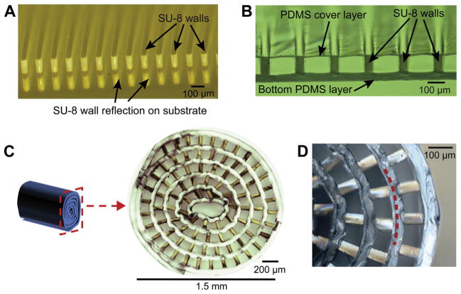
Microchannels and the rolled scaffold architecture. Complete Figure 3.
Srinivasan 2015 peripheral-nerve research scaffold; this figure shows the channel architecture.
Image credit & source
Srinivasan et al., Biomaterials (2015), Figure 3; © 2014 Elsevier Ltd. · Biomaterials · complete Figure 3 · © 2014 Elsevier Ltd. · source copyright retained.
Complete PMC source figure copied intact, byte for byte. All panels, annotations and scale bars retained; no cropping, re-encoding, upscaling or synthetic enhancement.
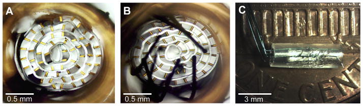
Microchannel interface assembly with integrated microwires, viewed from three sides. Complete Figure 9.
Representative wired scaffold assembly from the same 2015 peripheral-nerve study.
Image credit & source
Srinivasan et al., Biomaterials (2015), Figure 9; © 2014 Elsevier Ltd. · Biomaterials · complete Figure 9 · © 2014 Elsevier Ltd. · source copyright retained.
Complete PMC source figure copied intact, byte for byte. All panels, annotations and scale bars retained; no cropping, re-encoding, upscaling or synthetic enhancement.
- Code
- ?1R
- Reviewed
- 2026-02-06
- Interface
- pni
- Evidence stage
- research
Independent, source-linked catalog sheet. Not a manufacturer-issued datasheet, regulatory decision or instructions for clinical use. Human evidence does not establish approval. Source-specific restrictions, conflicts and missing specifications are retained below.
Microchannel-based regenerative scaffold interface (Srinivasan et al., 2015)
All rows follow the shared implant-device template. Measurements belong to the named study or configuration. A blank cell means the reviewed sources do not establish a value. Proposed use, terminal assays and continuously functioning implants are not treated as equivalent.
Identity
| Field | Value and source scope |
|---|---|
| Device | Srinivasan 2015 PDMS/SU-8 regenerative microchannel scaffold |
| Manufacturer | Academic fabrication in Srinivasan et al. |
| Interface class | Regenerative peripheral microchannel recording interface |
| Origin | Georgia Tech/Emory research team; design builds on prior microchannel work |
| First demonstrated | 2015 journal report, online December 2014; earliest family demonstration not established |
| First human implant | Reviewed study is rat preclinical work |
| Species studied | Rat sciatic amputee model |
| Regulatory status | Preclinical research |
| Function | Scaffold-guided regeneration and terminal neural recording after chronic implantation |
| Target tissue | Transected sciatic axons without distal reinnervation targets |
Geometry and architecture
| Field | Value and source scope |
|---|---|
| Interface type | Regenerative microchannels with integrated microwires |
| Array layout | PDMS top/bottom layers with SU-8 channel walls; scaffold rolled before implantation |
| Electrode count | Integrated microwire count not reported in audited publisher excerpts; number of channels is not assumed to equal electrodes |
| Pitch | 100 x 100 µm channel cross section; 20 x 100 µm walls in fabrication excerpt. Contact pitch not reported |
| Electrode lengths | 3 mm microchannel length in design discussion; not penetrating electrode length |
| Shank width and thickness | 40 µm base PDMS, 100 µm SU-8 layer, fabrication excerpt; total rolled scaffold dimensions not reported |
| Tip and exposed site geometry | Microwires integrated into bottom PDMS; exposed wire geometry not reported in audited excerpts |
| Contact coating | |
| Insulation | PDMS and SU-8 scaffold; microwire insulation not reported |
| Insertion method | Regenerating transected axons enter rolled microchannel scaffold in sciatic amputee model |
| Anchoring and fixation |
Electrode and channel physics
| Field | Value and source scope |
|---|---|
| Exposed site area | |
| Electrode material | Microwire conductor material not reported in audited excerpts; fabrication substrate Ti/Au anti-adhesion layers are not asserted as electrode material |
| Impedance (with measurement frequency) | |
| Noise floor or SNR | |
| Recording modality | Spontaneous, sensory-evoked and electrically evoked single-/multi-unit action potentials at five-month terminal experiment |
| Sampling rate | |
| Stimulation capability | Electrically evoked signals recorded; direct stimulation through integrated scaffold wires not established by audited excerpts |
| Charge injection limit | |
| Reference and ground |
Tissue interface and bioresponse
| Field | Value and source scope |
|---|---|
| Target tissue | Regenerating peripheral axons, Schwann cells and fibroblasts inside microchannels |
| Insertion trauma and BBB disruption | BBB: not applicable; model intentionally transects/amputates nerve |
| Vascular disruption risk | |
| Micromotion sensitivity | |
| Gliosis and encapsulation | Publisher abstract reports organized axons, endoneurial tubes and myelination distal to scaffold, without disorganized growth characteristic of neuroma; not a zero-neuroma clinical rate |
| Neuron loss near sites | |
| Foreign-body response mitigation | Biocompatible PDMS/SU-8 construction tested in vitro and in vivo; response not generalized to all packaging |
| Typical failure modes | Quantitative failure modes/rates not reported in audited excerpts; channel-clogging is not asserted as observed |
System architecture
| Field | Value and source scope |
|---|---|
| Onboard electronics | Passive microwires integrated in scaffold; active onboard electronics not reported |
| Data path | External terminal electrophysiology after chronic scaffold implant; chronic wiring/packaging not demonstrated in this study |
| Telemetry bandwidth | Not applicable to described terminal wired acquisition |
| Sampling rate | |
| Power | External electrophysiology equipment at terminal test |
| Thermal management | |
| Packaging and hermeticity | PDMS/SU-8 scaffold, no validated hermetic implanted electronic package |
| MRI compatibility | |
| Surgical complexity | Invasive regenerative nerve model with no distal target |
| Output connectors |
Performance envelope
| Field | Value and source scope |
|---|---|
| Acute yield | |
| Chronic yield | All devices in described terminal study recorded neural activity after five months; device count and per-contact yield not reported in audited abstract |
| Stability over time | Five-month endpoint recording, not five months of continuous recordings |
| Longevity | Five months implanted before terminal recording; maximum lifetime not reported |
| Revision and explant experience | Terminal testing; clinical revision experience not applicable |
| Adverse events | Clinical safety not reported; morphology findings not proof of absence of all complications |
| Notable demonstrations | Single-/multi-unit activity recorded from chronically implanted microchannels in amputee-model nerve |
Clinical and preclinical evidence
| Field | Value and source scope |
|---|---|
| Human subjects | Not applicable to reviewed preclinical study |
| Preclinical cohort | Rat sciatic amputee model; total and electrode-bearing subgroup counts not reported in audited publisher excerpts |
| Follow-up duration | Five-month terminal neural recording experiment |
| Indications | Preclinical prosthetic peripheral nerve interface after amputation |
| Trials and registries | Human registry not applicable |
| Primary outcomes | Cytocompatibility, organized regeneration and terminal electrophysiology |
| Key limitations | Publisher excerpts lack full electrode/connector details; five-month endpoint does not establish continuous chronic recording or human efficacy |
Engineering tradeoffs
| Field | Value and source scope |
|---|---|
| Strengths | Channels organize regeneration and permit terminal neural recordings after chronic implant |
| Limitations | Transection/amputee model and unresolved chronic wiring/packaging |
| Scaling constraints | Microchannel geometry, integrated wiring and rolling/assembly; large-electrode-count interface remains a stated goal |
References
- Srinivasan et al. 2015. Microchannel-based regenerative scaffold for chronic peripheral nerve interfacing in amputees. Publisher abstract and available fabrication/results excerpts; missing hardware details remain not reported.