Neuropixels 1.0 recording probe
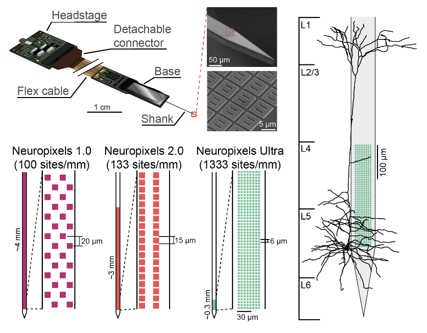
Neuropixels architecture at a glance: the assembled probe, microscopic contacts, and site layouts for 1.0, 2.0 and Ultra, alongside a cortical neuron.
The assembly and electron micrographs show Neuropixels Ultra. The contact-layout comparison provides context for the Neuropixels 1.0 specifications below.
Image credit & source
Ye and colleagues, Neuropixels Ultra study; architecture overview supplied by the site owner · Neuropixels Ultra · primary study · User-supplied image; source copyright retained.
Supplied image copied intact, byte for byte. All labels, panels, scale bars and embedded metadata retained.
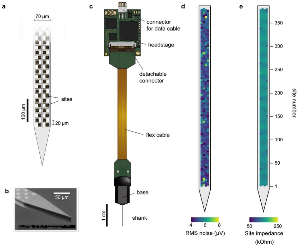
Neuropixels 1.0 in detail: recording sites and tip micrograph, the full probe and headstage, and site-by-site noise and impedance maps.
Original 2017 research probe shown in Jun and colleagues’ Figure 1. Labels and scale bars retain the original paper’s geometry.
Image credit & source
Jun et al., Nature (2017), Figure 1; image supplied by the site owner · Nature · Neuropixels 1.0 study · User-supplied image; source copyright retained.
Supplied image copied intact, byte for byte. All labels, panels, scale bars and embedded metadata retained.
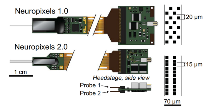
Neuropixels 1.0 and 2.0 compared: probe and headstage size, shank arrangement, and recording-site pitch.
The four-shank 2.0 design is the 2021 alpha research hardware. The comparison is a source illustration of two generations.
Image credit & source
Steinmetz et al., Science (2021), Figure 1A; image supplied by the site owner · Science · Neuropixels 2.0 study · User-supplied image; source copyright retained.
Supplied image copied intact, byte for byte. All labels, panels, scale bars and embedded metadata retained.
- Code
- L1R
- Reviewed
- 2026-10-07
- Interface
- intracortical
- Evidence stage
- research
Independent, source-linked catalog sheet. Not a manufacturer-issued datasheet, regulatory decision or instructions for clinical use. Human evidence does not establish approval. Source-specific restrictions, conflicts and missing specifications are retained below.
Neuropixels
NXT / NP3.0 is the latest announced generation, still a prototype. Opto, Ultra and NHP solve different problems and remain separate branches.
Highlighted family sheet: Neuropixels NXT / NP3.0 prototype →
Version history and parallel configurations (7)
- Neuropixels NXT / NP3.0 prototype Prototype: 1,536 channels, with up to 912 assigned to one shank. Purchase availability is a forecast.
- Neuropixels Opto photonic prototype Optical branch: electrical recording plus waveguides and 28 optical emitters.
- Neuropixels 2.0 Quad Base probe Quad Base: four shanks, 1,536 simultaneous channels, 384 per shank.
- Neuropixels Ultra Ultra: 6,144 small, densely packed sites, with 384 simultaneous recording channels.
- Neuropixels 1.0 NHP long-shank probe NHP: 45-mm shank, thicker mechanics and 4,416 selectable sites for deep primate access.
- Neuropixels 2.0 alpha single/four-shank probes 2.0 alpha: aligned dense sites, one/four-shank options and 384 channels per probe.
- Neuropixels 1.0 recording probe 1.0 baseline: 960 selectable sites and 384 simultaneous channels.
Interactive engineering reference
Neuropixels 1.0 — 3D Engineering Model
Explore the connector, gold interconnects, silicon shank, chisel tip and porous electrode detail. Switch between the drawing-matched study and reconstructed reference assembly inside the original viewer.
Open full-screen 3D viewer ↗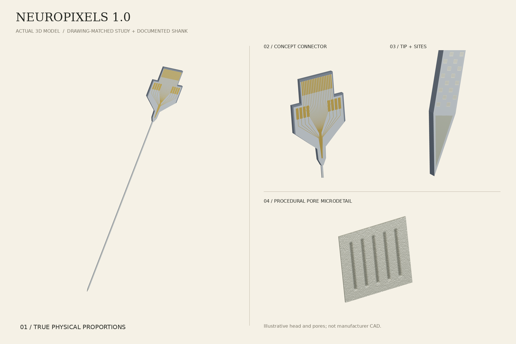
Nominal shank dimensions and 960 recording sites are documented. Connector routing, component placement and pore morphology include reconstructions; the drawing-matched head is a concept. Reference models — not for fabrication.
Models, drawings and source
Neuropixels 1.0 recording probe
This entry is the single-shank Neuropixels 1.0 recording probe, not every member of the family. Ultra and NHP have separate hardware entries. Neuropixels 2.0 has different site geometry and single/four-shank configurations; its counts must not be assigned to 1.0.
Identity
| Field | Value and source scope |
|---|---|
| Device | Neuropixels 1.0, single-shank recording probe [1, 2] |
| Manufacturer | imec, with HHMI Janelia, Allen Institute and UCL in the 2017 collaboration [2] |
| Interface class | Intracortical, penetrating silicon shank |
| Origin | imec with HHMI Janelia, Allen Institute and UCL collaboration [2] |
| First demonstrated | 2017 publication (Jun et al.) [2, 3] |
| First human implant | None reported; research-only device |
| Species studied | Rodent and other research animals; research-only device [2] |
| Regulatory status | Research tool; no clinical approval claimed |
| Function | Recording only [1] |
| Target tissue | Cortical and deep brain structures along the shank |
Geometry and architecture
| Field | Value and source scope |
|---|---|
| Interface type | Penetrating single shank |
| Array layout | Checkerboard with four column positions, two sites per row [1] |
| Electrode count | 960 selectable physical sites, 384 simultaneous channels [1] |
| Pitch | 16 µm across column positions, 20 µm between rows [1] |
| Electrode lengths | Shank 10 mm long [1] |
| Shank width and thickness | 70 µm wide; 20 µm thick in the 2017 paper, 24 µm in the later datasheet; both retained [1, 2] |
| Tip and exposed site geometry | 12 x 12 µm titanium nitride contacts [1]; the model tip outline and 200 µm first-row offset are approximations, not manufacturer drawings |
| Contact coating | Titanium nitride [1] |
| Insulation | |
| Insertion method | Stereotaxic placement in research surgery |
| Anchoring and fixation | Skull or headstage fixation in chronic preparations; configuration dependent |
Electrode and channel physics
| Field | Value and source scope |
|---|---|
| Exposed site area | 144 µm² nominal from 12 x 12 µm contacts [1] |
| Electrode material | Titanium nitride contacts [1] |
| Impedance (with measurement frequency) | Earlier mixed-generation values were removed |
| Noise floor or SNR | Not given as a single figure here; the 2017 paper reports low-noise performance [2] |
| Recording modality | Action potential and local field potential bands [1] |
| Sampling rate | 30 kHz AP / 2.5 kHz LFP, datasheet [1] |
| Stimulation capability | Not applicable; recording-only probe |
| Charge injection limit | Not applicable |
| Reference and ground | Configuration dependent on the headstage setup |
Tissue interface and bioresponse
| Field | Value and source scope |
|---|---|
| Target tissue | Brain parenchyma along the insertion track |
| Insertion trauma and BBB disruption | Inherent to penetrating silicon shanks; quantitative values not reported |
| Vascular disruption risk | |
| Micromotion sensitivity | Rigid shank; qualitative concern, quantitative data not extracted here |
| Gliosis and encapsulation | Not extracted in this sheet |
| Neuron loss near sites | Not extracted in this sheet |
| Foreign-body response mitigation | |
| Typical failure modes | Recording failures documented in the 2017 study; see the linked application entry [2] |
System architecture
| Field | Value and source scope |
|---|---|
| Onboard electronics | On-base signal conditioning, amplification, multiplexing and digitization; the preferred design was passive switched, not one amplifier per site [2] |
| Data path | Wired external headstage |
| Telemetry bandwidth | Wired; configuration dependent |
| Sampling rate | 30 kHz AP / 2.5 kHz LFP, datasheet [1] |
| Power | External |
| Thermal management | Low dissipation; specifics not reported here |
| Packaging and hermeticity | Non-hermetic research probe |
| MRI compatibility | |
| Surgical complexity | Stereotaxic research surgery |
| Output connectors |
Performance envelope
| Field | Value and source scope |
|---|---|
| Acute yield | Large simultaneous neuronal populations reported in the 2017 paper; see the application entry [2] |
| Chronic yield | Durability limits and failures documented in the linked 2017 study entry [2] |
| Stability over time | Weeks-scale research recordings; not a lifetime implant |
| Longevity | Not specified as an implant-lifetime figure |
| Revision and explant experience | Not applicable to research use |
| Adverse events | Not applicable as a clinical device |
| Notable demonstrations | Two-probe population recordings in the 2017 study [2] |
Clinical and preclinical evidence
| Field | Value and source scope |
|---|---|
| Human subjects | None; research-only device |
| Preclinical cohort | 2017 rodent study and follow-on literature [2] |
| Follow-up duration | |
| Indications | Neuroscience research |
| Trials and registries | None |
| Primary outcomes | |
| Key limitations | Research-only; chronic durability limited per the linked study |
Engineering tradeoffs
| Field | Value and source scope |
|---|---|
| Strengths | Very high site density on one shank, on-probe conditioning and digitization, mature research ecosystem |
| Limitations | 384 simultaneous channels against 960 sites, rigid tethered shank, research-only packaging |
| Scaling constraints | Multiplexing and readout bandwidth, shank cross-section versus tissue displacement, chronic packaging |
Manufacturer specification
| Feature | Neuropixels 1.0 datasheet |
|---|---|
| Available recording sites | 960 |
| Simultaneous recording channels | 384 |
| Shank | 10 mm long, 70 µm wide, 24 µm thick |
| Recording contacts | 12 × 12 µm titanium nitride |
| Contact arrangement | Checkerboard, four column positions; two sites per row |
| Pitch | 16 µm across column positions, 20 µm between rows |
| Bands | Action potential and local field potential |
| AP / LFP sample rates | 30 kHz / 2.5 kHz |
A site is a physical electrode. A channel is a signal-processing and readout path. Selecting 384 sites does not turn the other 576 sites into simultaneous channels. The external wired headstage and acquisition system remain part of the setup.
Original paper versus later datasheet
Jun and colleagues’ 2017 paper reports a 10 mm shank with 70 × 20 µm cross-section. The manufacturer datasheet gives 70 × 24 µm. Both figures are retained as source-specific descriptions, not averaged or silently treated as interchangeable.
The viewer uses the datasheet’s 24 µm shank thickness, 960 sites, 12 µm square contacts and checkerboard pitch. Its tip outline and 200 µm first-row offset are approximations. It omits base electronics, headstage and acquisition mapping; contact IDs are geometric labels, not actual channel assignments.
Recording circuitry
The original development compared passive, active, switched and active-switched designs. The preferred design was passive switched. The paper’s on-base signal conditioning, amplification, multiplexing and digitization must not be simplified to a universal claim that every 1.0 electrode has an on-site amplifier.
Demonstrated recordings and failures
The 2017 rodent study and durability limits separate two-probe population recordings from chronic event-rate results. The paper reports failures as well as stable recordings. These experiments do not establish a permanent human implant or clinical BCI indication.
References
Models and reference visualizations
Neuropixels 1.0 · recording shank
Electrode-bearing structure · dimensions in millimeters
Loading 3D viewer…
Model fidelity. Neuropixels 1.0 only; research recording probe. True-scale shank and sites. The 175 µm in-plane tip taper follows the manufacturer datasheet; the 200 µm tip-to-first-row offset is approximate. Silicon and porous TiN have distinct neutral finishes; colors and roughness are visual approximations. All 960 sites are shown; the selectable 384-channel mapping is not modeled. External base electronics, flex cable and headstage are omitted. Use Tip detail to see contacts at true scale.
Color, transparency and surface finish approximate the references. Use Highlight contacts to inspect electrode locations.
Sources & reference images
Dimensions come from the linked specifications; images guide shape only. Reference images remain on their owners’ websites.
- Neuropixels 1.0 datasheet — 175 µm tip length and shank dimensions →
- Neuropixels — 1.0 probe specifications and gallery →
- Steinmetz et al. (2021) — NP 1.0 / 2.0 geometry comparison →
- Allen Institute — contact geometry →
- Neuropixels Central — 1.0 product photograph →
JSON includes physical site coordinates, unknown channel mappings and approximation notes. GLB uses meters. These models do not predict recording or stimulation performance.
What a Neuropixels probe records
One Neuropixels 1.0 probe, spike-sorted, from a mouse performing the IBL visual decision task. Each row is a sorted unit, placed at the depth it was detected along the 3.84 mm of recording sites. These are measured recordings, not simulated signals.
Loading the recording excerpt...
Source, license & limitations
International Brain Laboratory et al. (2023). A brain-wide map of neural activity during complex behaviour. Public data via OpenAlyx, released under CC BY 4.0 (AWS Open Data Registry listing). Session ebce500b-c530-47de-8cb1-963c552703ea, probe00, pykilosort spike sorting, mouse MFD_09, recorded October 19, 2023 at the Churchland lab.
Only units labeled good by the IBL quality criteria are shown. The raster is a 20-second excerpt (trials 151-155); the stimulus-aligned averages use every trial in the session. Spike depths are the sorter's estimates, not exact anatomical positions, and region labels come from histology-aligned channel locations. Sorted units are an estimate of single neurons.
This is a mouse recording, not a human implant, and the viewer does not run a decoder. Neuropixels probes are acute research tools; the 3D model above is the probe's physical geometry, and unit depth here is not mapped contact by contact onto it.