Neuralink N1 (flexible-thread implant)
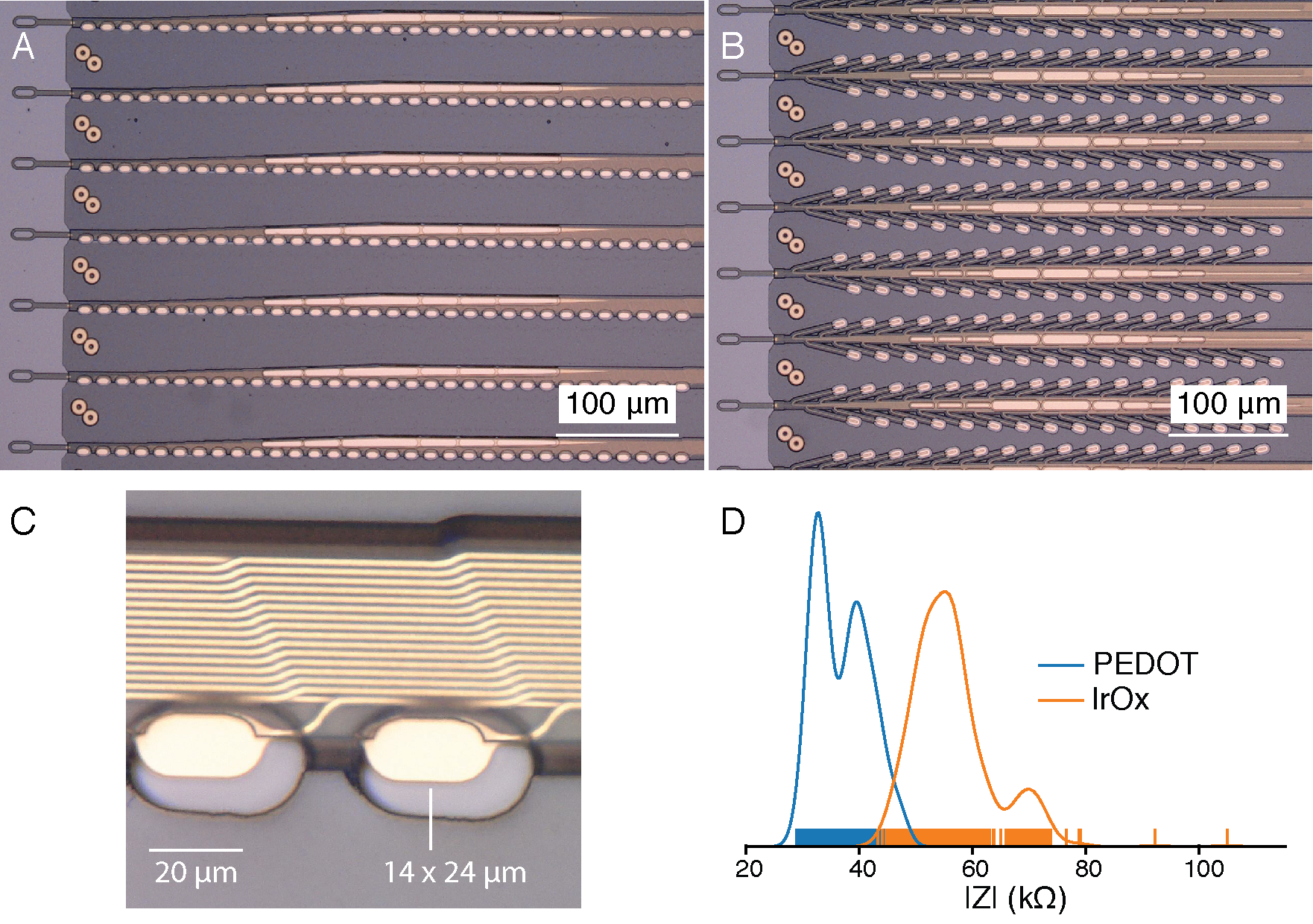
Neuralink’s 2019 research thread architecture: two polymer probe designs, electrode contacts and impedance measurements. Historical reference, not the current N1 implant.
Historical 2019 wired research platform, not the current wireless N1 implant. N1 specifications below describe a later generation.
Image credit & source
Elon Musk and Neuralink, 2019 research white paper, published in JMIR 21(10):e16194 · Neuralink · 2019 research white paper · CC BY-ND 4.0.
Complete figure recovered at its native embedded resolution from the 2019 white paper. Transparency composited on the paper’s white background; no cropping, upscaling, retouching or panel removal. The recovered PNG is copied intact for both display and enlargement.
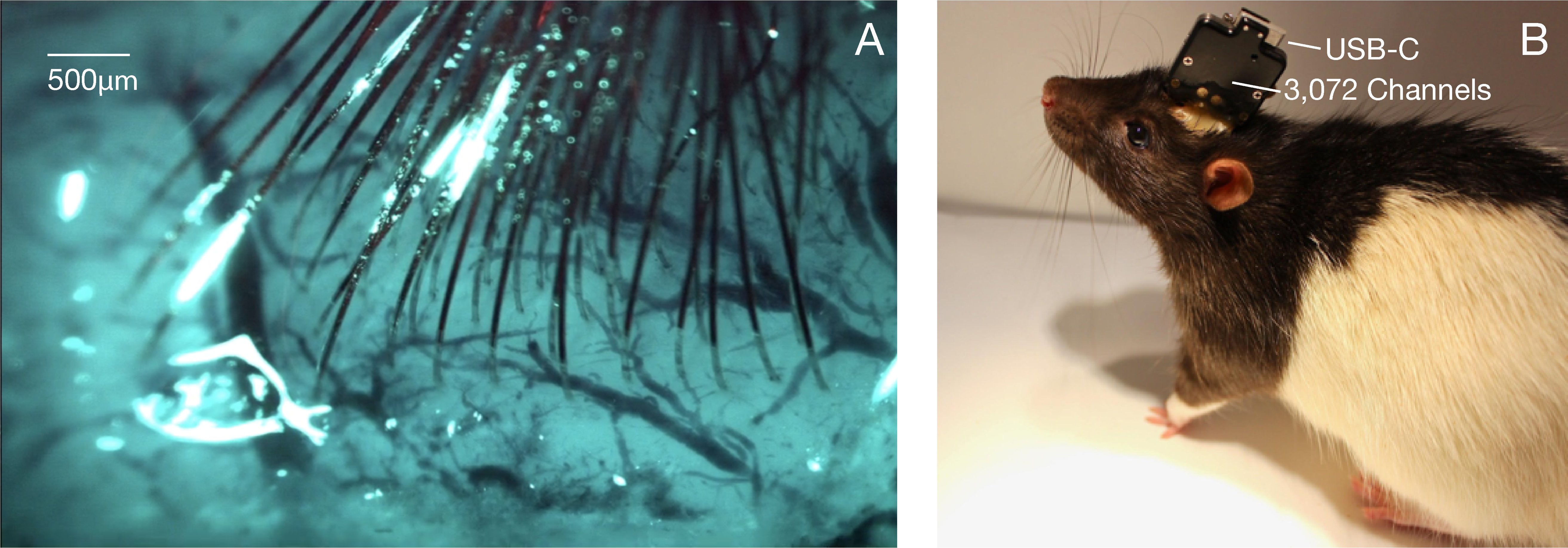
Threads implanted in rat cortex, with the original 500 µm scale, beside the wired research recording package. Complete 2019 Figure 6; this does not depict the current N1 implant.
Historical 2019 wired research platform, not the current wireless N1 implant. N1 specifications below describe a later generation.
Image credit & source
Elon Musk and Neuralink, 2019 research white paper, published in JMIR 21(10):e16194 · Neuralink · 2019 research white paper · CC BY-ND 4.0.
Complete figure recovered at its native embedded resolution from the 2019 white paper. Transparency composited on the paper’s white background; no cropping, upscaling, retouching or panel removal. The recovered PNG is copied intact for both display and enlargement.
- Code
- C3R
- Reviewed
- 2026-10-07
- Interface
- intracortical
- Evidence stage
- human
Independent, source-linked catalog sheet. Not a manufacturer-issued datasheet, regulatory decision or instructions for clinical use. Human evidence does not establish approval. Source-specific restrictions, conflicts and missing specifications are retained below.
Neuralink N1 Implant
N1 combines independently placed flexible electrode threads with a skull-mounted enclosure containing electronics, a rechargeable battery and wireless telemetry. The R1 robot inserts the threads; an external application decodes the recorded activity into computer actions. [1]
Which N1 is modeled?
This is the 64-thread, 16-sites-per-thread configuration described in 2024, totaling 1,024 physical sites. [1, 2] The dated name matters: UCLH’s July 2025 GB-PRIME description instead specifies 128 threads with 8 electrodes each. [3] Both total 1,024 sites, but they have different physical layouts. The newer description is linked for comparison and is not silently folded into the 2024 model.
Geometry and evidence
| Parameter | Evidence for the selected revision | Treatment in the model |
|---|---|---|
| Threads / electrodes | 64 / 1,024 [1] | 64 separately drawn ribbons, 16 sites each |
| Along-thread site pitch | 200 µm, reported by DJ Seo in 2024 [2] | 200 µm center to center |
| Thread width | 16–84 µm in the same interview [2] | Linear taper between those limits; progression is illustrative |
| Thread thickness | Two 2 µm polymer layers and a 0.4 µm metal stack [2] | 4.4 µm total derived from the described stack |
| Enclosure | Approximately quarter-sized and 9 mm thick [2] | Nominal 24 mm diameter × 9 mm thickness |
| Contact shape and exposed area | 12 × 20 µm visual markers; exposed area is exported as unknown | |
| Thread placement | Independently placed by the surgical robot [1] | Unfurled display fan, not a cortical insertion map |
Using the 3D reference
The model shows the enclosure envelope and all 64 flexible ribbons in a schematic unfurled pose. Thread detail shows a terminal recording section; Contact detail reveals the tiny site markers. Both change the camera only. Gold is a visual highlight, not a claim about the exposed contact’s actual appearance.
Fan length, thread-to-thread spacing, bends, taper progression and enclosure trim are reconstruction choices. Internal chips, the charging coil, surgical insertion loops and individual metal traces are omitted. ClinicalTrials.gov describes the N1 as a skull-mounted, wireless, rechargeable implant [4], so a coil exists, but no cited source gives its size or position and none is drawn. No patient-specific trajectory or cortical insertion depth is encoded in this display pose.
JSON exports use millimeters and identify both thread and site index. Each physical site has a unique geometric ID; acquisition channel assignments and contact areas stay null. GLB uses meters and includes the full assembly even when viewing contacts only. Transforming the display fan into an implanted configuration requires independently specified trajectories and a validated electrical model.
Interface and operation
The engineering interview describes a polyimide-insulated thin-film metal stack and iridium-oxide recording sites, with electrodes distributed along the thread rather than only at its endpoint. It also discusses stimulation capability; that does not establish clinical efficacy or stimulation limits for this geometric model. [2]
Inductive charging and wireless data allow the implant to operate without a percutaneous connector. [1] The relevant human programs are investigational studies. UCLH describes the GB-PRIME study as evaluating safety and functionality, with an initial study period and longer follow-up. [3]
Identity
| Field | Value and source scope |
|---|---|
| Device | N1 implant, flexible-thread intracortical BCI |
| Manufacturer | Neuralink |
| Interface class | Intracortical, penetrating flexible threads |
| Origin | Neuralink; first human participant reported in company updates from 2024 [1] |
| First demonstrated | 2024 company update for the 64-thread, 16-site description [1, 2]; the July 2025 UCLH description gives 128 threads of 8 electrodes [3] |
| First human implant | First PRIME participant reported in company updates beginning 2024 [1] |
| Species studied | Human (investigational) and preclinical animals; preclinical detail not reported here |
| Regulatory status | Investigational: PRIME (NCT06429735) [4] and GB-PRIME [3]; not commercially approved |
| Function | Recording; stimulation capability discussed in the engineering interview [2] |
| Target tissue | Motor cortex in the reported human program |
Geometry and architecture
| Field | Value and source scope |
|---|---|
| Interface type | Penetrating flexible polymer threads with a skull-mounted enclosure |
| Array layout | 64 threads x 16 sites (2024 description) [1, 2]; the 128 x 8 variant is documented separately [3] |
| Electrode count | 1,024 physical sites in both descriptions [1, 3]; simultaneous channel count not reported |
| Pitch | 200 µm along the thread, per the 2024 interview [2] |
| Electrode lengths | Thread reach and insertion depth are patient-specific and not published |
| Shank width and thickness | 16-84 µm width; thickness about 4.4 µm derived from two 2 µm polymer layers and a 0.4 µm metal stack [2] |
| Tip and exposed site geometry | Contact shape and exposed area not reported; model markers are illustrative |
| Contact coating | Iridium oxide recording sites on a thin-film metal stack [2] |
| Insulation | Polyimide over a thin-film metal stack [2] |
| Insertion method | R1 surgical robot with a needle that grasps, inserts and releases threads [1]; company page describes five cameras and an OCT system |
| Anchoring and fixation | Skull-mounted enclosure [1, 4]; thread anchoring in tissue not reported |
Electrode and channel physics
| Field | Value and source scope |
|---|---|
| Exposed site area | Exported as unknown |
| Electrode material | Iridium-oxide recording sites on a thin-film metal stack [2] |
| Impedance (with measurement frequency) | |
| Noise floor or SNR | |
| Recording modality | Recording of neural activity near neurons to detect action potentials per the company page; bands not reported |
| Sampling rate | |
| Stimulation capability | Discussed publicly [2]; parameters and limits not reported |
| Charge injection limit | |
| Reference and ground |
Tissue interface and bioresponse
| Field | Value and source scope |
|---|---|
| Target tissue | Cerebral cortex |
| Insertion trauma and BBB disruption | Threads are placed to avoid vasculature per company descriptions [1]; quantitative trauma data not reported |
| Vascular disruption risk | Threads placed to avoid vasculature per company descriptions [1]; quantitative data not reported |
| Micromotion sensitivity | Flexible threads are designed to move with tissue [1]; independent quantitative data not reported |
| Gliosis and encapsulation | |
| Neuron loss near sites | |
| Foreign-body response mitigation | Thread flexibility and small cross-section are the design approach; measured outcomes not reported |
| Typical failure modes | Thread retraction after implantation was reported for the first participant in company updates [1] |
System architecture
| Field | Value and source scope |
|---|---|
| Onboard electronics | Enclosure contains electronics for recording, processing and wireless telemetry; custom low-power chips per the company page [1] |
| Data path | Wireless to the Neuralink Application, which decodes the data stream into actions; no percutaneous connector [1] |
| Telemetry bandwidth | |
| Sampling rate | |
| Power | Small rechargeable battery charged wirelessly with an inductive charger [1, 4] |
| Thermal management | |
| Packaging and hermeticity | Hermetically sealed biocompatible enclosure per the company page; hermeticity specifications not reported |
| MRI compatibility | |
| Surgical complexity | R1 robot insertion with skull-mounted enclosure placement [1] |
| Output connectors | No percutaneous connector [1]; wireless link details not reported |
Performance envelope
| Field | Value and source scope |
|---|---|
| Acute yield | |
| Chronic yield | Company reports cursor and device control in study participants [1]; independent datasets not reported |
| Stability over time | Thread retraction affected early recordings in the first participant [1]; long-term stability not reported |
| Longevity | Battery and packaging lifetime not publicly specified |
| Revision and explant experience | |
| Adverse events | No independently audited rates |
| Notable demonstrations | Company-reported cursor control and computer use by study participants [1] |
Clinical and preclinical evidence
| Field | Value and source scope |
|---|---|
| Human subjects | PRIME and GB-PRIME investigational studies; company-reported participant counts change over time and are not pinned here |
| Preclinical cohort | Preclinical cohort details not reported here |
| Follow-up duration | Initial study period with longer follow-up per UCLH [3] |
| Indications | Severe motor impairment and paralysis |
| Trials and registries | NCT06429735 (PRIME) [4]; GB-PRIME [3] |
| Primary outcomes | Safety and functionality [3, 4] |
| Key limitations | Evidence is largely company statements and interviews; peer-reviewed human data remain sparse |
Engineering tradeoffs
| Field | Value and source scope |
|---|---|
| Strengths | High site count, fully implanted wireless operation, flexible threads, robotic insertion, no percutaneous connector |
| Limitations | Sparse peer-reviewed data, reported thread retraction, dependence on a novel surgical robot, unpublished longevity |
| Scaling constraints | Surgical throughput, hermetic packaging, telemetry bandwidth, power and heat, chronic tissue response |
References
- Neuralink — PRIME Study Progress Update; April 12, 2024. Includes the manufacturer’s exploded N1 reference image.
- DJ Seo — Neuralink engineering interview, Lex Fridman podcast #438 (2024). See 02:03:33 for electrode layout, 02:07:03 for enclosure, 02:08:58 for width and 02:12:48 for the layer stack. These are public engineering descriptions, not released fabrication drawings.
- UCLH — GB-PRIME study description; July 31, 2025. Documents a separate 128 × 8 arrangement and investigational study status.
- ClinicalTrials.gov NCT06429735 — PRIME study. Describes the N1 Implant as skull-mounted, wireless and rechargeable.
The company technology page (neuralink.com/technology) describes a hermetically sealed biocompatible enclosure, a battery charged wirelessly by an inductive charger, custom low-power chips, and a surgical robot with five camera systems, an optical coherence tomography system and a needle that grasps, inserts and releases threads. Third-party device databases give figures such as a coin-sized 23 x 18 x 2 mm body and 200 Mbps raw data; those are not confirmed by Neuralink in the reviewed sources and are not used here.
Models and reference visualizations
Neuralink N1 · 64 × 16 · 2024 reference
Electrode-bearing structure · dimensions in millimeters
Loading 3D viewer…
Model fidelity. Named 64-thread configuration described in 2024, not every N1 generation. UCLH later describes 128 threads × 8 sites. The fan is an unimplanted display pose, not a cortical placement map. 24 mm diameter approximates the quarter-sized enclosure; rounded enclosure profile, fan length, taper progression, spacing, strain-relief tab and 12 × 20 µm site markers are illustrative. The pale smooth enclosure follows the 2024 exploded view; the tab spans the full reconstructed thread fan. Optical finishes are illustrative, and unsupported decorative concentric inlays are omitted. The 4.4 µm thickness is the sum of the interview’s 2 µm polymer + 0.4 µm metal + 2 µm polymer layers. ClinicalTrials.gov describes the implant as skull-mounted, wireless and rechargeable, so it has a charging coil, but no cited source gives dimensions sufficient to reconstruct its geometry, so no coil is drawn. UCLH describes the enclosure as the size of a ten-pence coin (about 24 mm), consistent with the modeled diameter. Actual contact shape/area, insertion loops, individual metal traces and internal electronics are unresolved. Camera detail views retain physical scale. Site IDs are geometric labels, not acquisition channel assignments.
Color, transparency and surface finish approximate the references. Use Highlight contacts to inspect electrode locations.
Sources & reference images
Dimensions come from the linked specifications; images guide shape only. Reference images remain on their owners’ websites.
- Neuralink — PRIME Study Progress Update (April 2024): 64 threads, 1,024 electrodes →
- DJ Seo, Neuralink engineering interview (2024), 02:03–02:15 — pitch, thread stack and enclosure →
- ClinicalTrials.gov NCT06429735 — PRIME: N1 described as skull-mounted, wireless, rechargeable →
- UCLH (2025) — separately described 128 × 8 trial configuration →
- Neuralink — N1 exploded view from the 2024 PRIME update →
JSON includes physical site coordinates, unknown channel mappings and approximation notes. GLB uses meters. These models do not predict recording or stimulation performance.