ActiGait implanted drop-foot stimulator (common peroneal nerve multi-contact cuff)
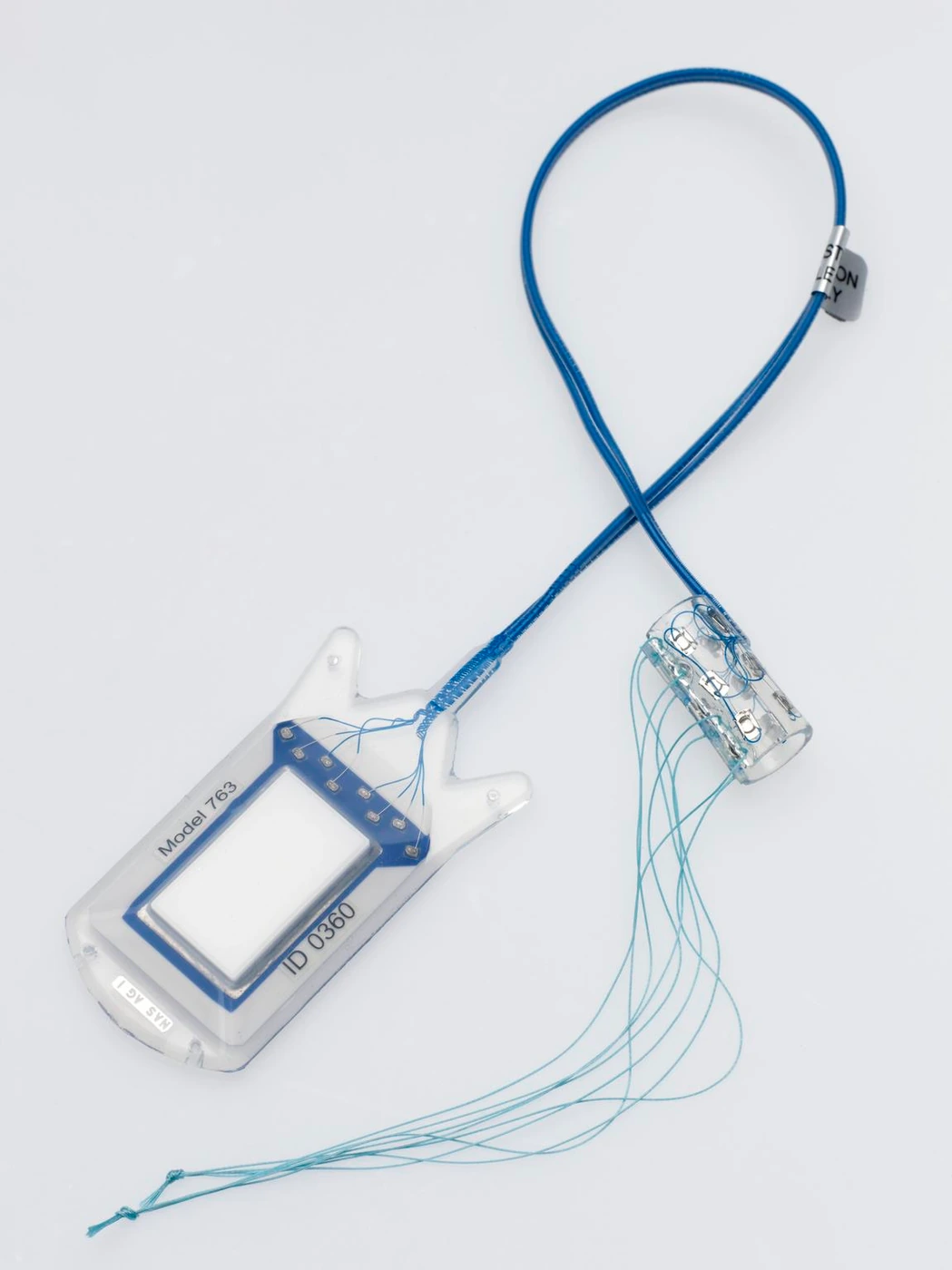
ActiGait receiver, cable and nerve cuff, circa 2010 demonstration sample. Actual museum-object photograph; the sample is marked for demonstration use.
Museum dates the object to circa 2010. Demonstration sample, not an explanted patient device; later clinical configurations may differ.
Scale: No calibrated scale bar; museum dimensions describe the overall specimen and are not a contact-pitch calibration.
Historical transparent demonstration sample exposes cuff wiring and contacts. Exact inner-cuff dimensions should use configuration-specific technical sources.
Image credit & source
Science Museum Group Collection © The Board of Trustees of the Science Museum; Otto Bock Healthcare Products, object 2021-634 · Science Museum Group · ActiGait implant · CC BY-NC-SA 4.0.
Photograph resized and encoded as WebP; no retouching or generative changes.
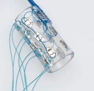
ActiGait demonstration cuff close-up: transparent body, contact plates and individual lead routing. No scale bar.
Museum dates the object to circa 2010. Demonstration sample, not an explanted patient device; later clinical configurations may differ.
Scale: No calibrated scale bar; museum dimensions describe the overall specimen and are not a contact-pitch calibration.
Historical transparent demonstration sample exposes cuff wiring and contacts. Exact inner-cuff dimensions should use configuration-specific technical sources.
Image credit & source
Science Museum Group Collection © The Board of Trustees of the Science Museum; Otto Bock Healthcare Products, object 2021-634 · Science Museum Group · cuff detail · CC BY-NC-SA 4.0.
Nerve cuff mechanically cropped from the museum photograph; resized and encoded as WebP. No retouching or generative changes.
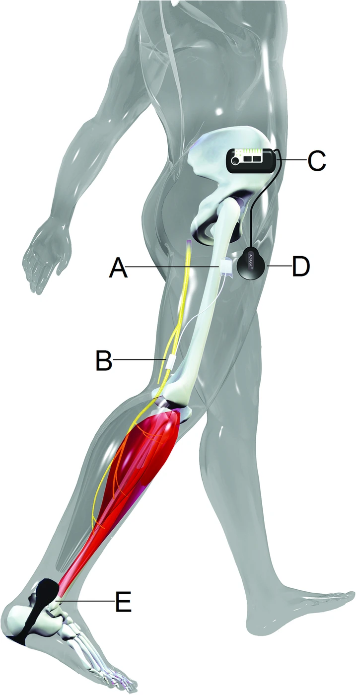
ActiGait implantation and system layout. A: implanted stimulator, B: peroneal nerve cuff; C–E: external controller, antenna and heel switch. Source illustration, not a hardware photograph.
ActiGait system depiction used in the 2019 clinical report; current availability or configuration is not inferred.
Scale: Illustration without a calibrated scale bar.
Placement diagram only. Cuff electrode contacts and exact dimensions are not resolved; it cannot support an as-built cuff blueprint.
Image credit & source
Berenpas et al., Journal of NeuroEngineering and Rehabilitation (2019), Fig. 2 · Berenpas 2019 · Fig. 2 · CC BY 4.0 (article); third-party source image rights not separately established.
Complete source illustration retained; resized and encoded as WebP. No generative changes.
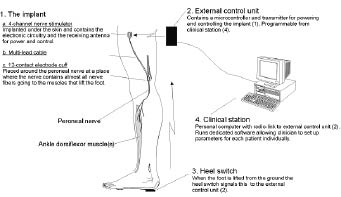
ActiGait system diagram from the Danish phase-II trial by Neurodan, Aalborg and Southampton investigators (2007). Historical illustration.
Historical phase-II ActiGait system illustration from investigators including Neurodan A/S, Denmark; low-resolution original.
Scale: Illustration without a calibrated scale bar.
Placement diagram only. Cuff electrode contacts and exact dimensions are not resolved; it cannot support an as-built cuff blueprint.
Image credit & source
Burridge, Haugland et al., Journal of Rehabilitation Medicine (2007), Fig. 1 · Burridge / Haugland 2007 · Fig. 1 · Copyright reserved; historical source figure reference.
Complete source illustration retained; resized and encoded as WebP. No generative changes.
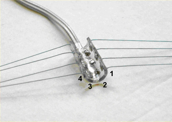
ActiGait cuff detail from the 2004 Haugland/Larsen/Burridge presentation. The four groups of tripolar electrodes are labeled 1–4 in the source.
Historical 2004 ActiGait configuration, sourced from Danish-hosted primary author presentation; not assumed identical to later clinical hardware.
Scale: No calibrated scale bar.
Four tripolar groups are visible. This historic presentation illustrates contact arrangement, but exact cuff diameter/contact pitch cannot be measured without a calibrated source.
Image credit & source
Haugland, Larsen, Burridge et al., ActiGait IFESS presentation (2004), slide 6 · Haugland et al. 2004 · cuff detail · Copyright reserved; historical author presentation reference.
Original embedded photograph extracted from presentation slide 6, resized and encoded as WebP; original contact labels retained. No generative changes.
- Code
- ?3?
- Reviewed
- 2026-02-06
- Interface
- pni
- Evidence stage
- human
Independent, source-linked catalog sheet. Not a manufacturer-issued datasheet, regulatory decision or instructions for clinical use. Human evidence does not establish approval. Source-specific restrictions, conflicts and missing specifications are retained below.
ActiGait implanted drop-foot stimulator (common peroneal nerve multi-contact cuff)
Every field follows the shared implant-device template. Values belong to a named configuration or study; not reported means the reviewed sources do not establish the value. Family-wide and deployment-specific evidence are kept separate.
Identity
| Field | Value and source scope |
|---|---|
| Device | ActiGait partly implantable drop-foot stimulation system |
| Manufacturer | ActiGait brand in Martin clinical studies; historical manufacturer not inferred from current ownership |
| Interface class | Extraneural peroneal nerve cuff with implanted stimulator and external gait control |
| Origin | Reviewed clinical studies in Dresden; not an invention-priority claim |
| First demonstrated | |
| First human implant | |
| Species studied | Human stroke and multiple sclerosis cohorts, Martin 2016/36-month study |
| Regulatory status | Clinical study evidence; present market availability and current authorization not reported |
| Function | Gait-triggered stimulation for dorsiflexion |
| Target tissue | Common peroneal nerve motor branch |
Geometry and architecture
| Field | Value and source scope |
|---|---|
| Interface type | Non-penetrating peripheral nerve cuff |
| Array layout | Four stimulation channels positioned at 90° around circumference, 36-month study |
| Electrode count | Four stimulation channels, 36-month study; physical contact construction not separately reported |
| Pitch | 90° angular channel arrangement; linear pitch not reported |
| Electrode lengths | Not applicable to cortical shanks; cuff length not reported |
| Shank width and thickness | Not applicable; cuff wall dimensions not reported |
| Tip and exposed site geometry | |
| Contact coating | |
| Insulation | |
| Insertion method | Two-incision surgery under general anesthesia; nerve exposed for 4 cm above tibial plateau, cuff placed around motor branch, 36-month study |
| Anchoring and fixation | Stimulator sutured to thigh muscle fascia, lead tunneled subcutaneously over biceps tendon; cuff closed around nerve, 36-month study |
Electrode and channel physics
| Field | Value and source scope |
|---|---|
| Exposed site area | |
| Electrode material | |
| Impedance (with measurement frequency) | |
| Noise floor or SNR | |
| Recording modality | Not applicable to described stimulation-only use |
| Sampling rate | Not applicable to passive cuff; heel-switch control is separate |
| Stimulation capability | Four selectable stimulation channels; patient-specific settings in Table 1 of 36-month study, not a universal safety envelope |
| Charge injection limit | |
| Reference and ground |
Tissue interface and bioresponse
| Field | Value and source scope |
|---|---|
| Target tissue | Peroneal nerve surface |
| Insertion trauma and BBB disruption | BBB: not applicable; nerve surgically exposed and cuffed. Martin 2016 reports nerve injury in two patients |
| Vascular disruption risk | |
| Micromotion sensitivity | |
| Gliosis and encapsulation | Peripheral interface; quantitative fibrosis/histology not reported in audited studies |
| Neuron loss near sites | |
| Foreign-body response mitigation | |
| Typical failure modes | Martin 2016: nerve injury requiring cuff repositioning and infection requiring system removal; later study also notes heel-switch malfunction |
System architecture
| Field | Value and source scope |
|---|---|
| Onboard electronics | Implanted stimulator body in thigh connected by cable to cuff |
| Data path | External heel switch sends radiofrequency signal to external control unit; antenna sends power/control to implanted stimulator, 36-month study |
| Telemetry bandwidth | |
| Sampling rate | Not applicable to described neural recording; stimulation timing patient-specific |
| Power | External control unit transfers energy via external antenna to implant; 36-month study |
| Thermal management | |
| Packaging and hermeticity | Subcutaneous stimulator and lead; hermetic qualification not reported |
| MRI compatibility | Preoperative MRI is not evidence of post-implant MR safety |
| Surgical complexity | General anesthesia, two incisions, nerve dissection and thigh stimulator placement |
| Output connectors | Internal electrode cable; external wireless heel-switch/control chain, connector dimensions not reported |
Performance envelope
| Field | Value and source scope |
|---|---|
| Acute yield | |
| Chronic yield | |
| Stability over time | 36-month study reports maintained gait benefit; not channel-yield or hardware reliability rate |
| Longevity | 36 months follow-up in later study; not maximum service life |
| Revision and explant experience | Martin 2016: two cuff repositionings for nerve injury and one system removal for wound infection among 27 patients |
| Adverse events | Martin 2016 also reports one delayed wound healing; later study notes heel-switch malfunction |
| Notable demonstrations | Martin 2016: 20 m walk time 33.9 to 17.9 seconds, six-minute distance 196 to 401 m; cohort averages, not guaranteed benefit |
Clinical and preclinical evidence
| Field | Value and source scope |
|---|---|
| Human subjects | 2016: 27 stroke patients; later study: 33 total, 27 stroke and six MS. Cohorts overlap, do not add as unique people |
| Preclinical cohort | |
| Follow-up duration | Later study evaluates baseline and 36 months |
| Indications | Central drop foot after stroke or MS with response to surface stimulation |
| Trials and registries | Prospective clinical studies; registry identifier not reported in audited sources |
| Primary outcomes | Gait speed, endurance, timed up-and-go and quality of life |
| Key limitations | Selected surface-stimulation responders; small uncontrolled cohorts; current product status not established |
Engineering tradeoffs
| Field | Value and source scope |
|---|---|
| Strengths | Selective gait-timed peroneal nerve stimulation with human follow-up |
| Limitations | Implant surgery and external heel-switch/antenna burden; revision and infection documented |
| Scaling constraints | Four-channel gait-focused system; no high-density scaling claim in reviewed studies |
References
- Martin et al. 2016. Restoration of ankle movements with the ActiGait implantable drop foot stimulator. Clinical abstract, 27-patient cohort.
- ActiGait 36-month clinical follow-up. Full device description, surgery and overlapping 33-patient cohort.