Hyperflexible Regenerative Sieve
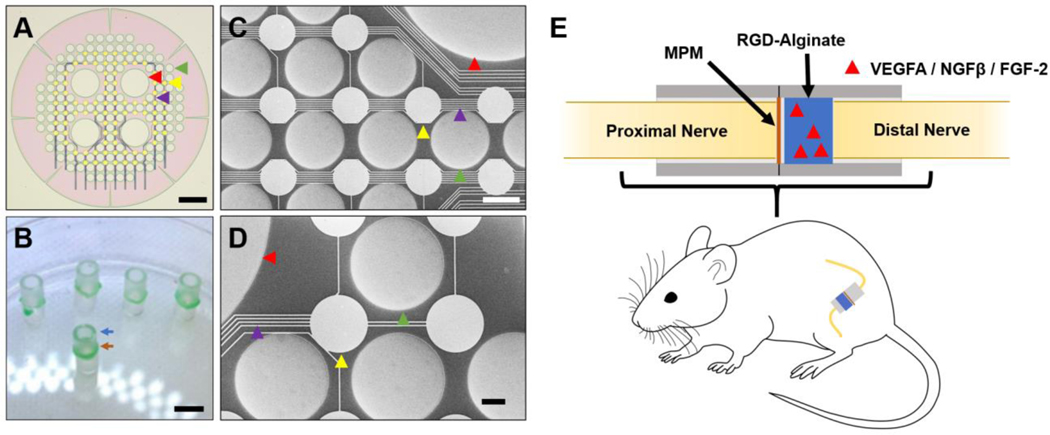
Redesigned mixed-porosity sieve, its assembly and microscopic contacts. Complete Figure 3.
2021 rat sciatic-nerve research configuration; distinct from the initial square-pore mesh in Figure 1.
Image credit & source
Veith et al., Biomaterials (2021), Figure 3; © Elsevier Ltd. · Biomaterials · complete Figure 3 · © 2021 Elsevier Ltd. · source copyright retained.
Complete PMC source figure copied intact, byte for byte. All panels, annotations and scale bars retained; no cropping, re-encoding, upscaling or synthetic enhancement.
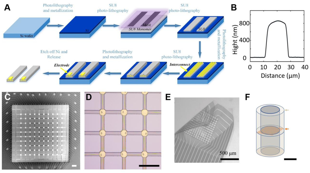
Initial square-pore sieve: fabrication, microscopy and the released flexible mesh. Complete Figure 1.
Initial 2021 design; its geometry and outcomes must be distinguished from the redesigned mesh in Figure 3.
Image credit & source
Veith et al., Biomaterials (2021), Figure 1; © Elsevier Ltd. · Biomaterials · complete Figure 1 · © 2021 Elsevier Ltd. · source copyright retained.
Complete PMC source figure copied intact, byte for byte. All panels, annotations and scale bars retained; no cropping, re-encoding, upscaling or synthetic enhancement.
- Code
- ?1R
- Reviewed
- 2026-02-06
- Interface
- pni
- Evidence stage
- research
Independent, source-linked catalog sheet. Not a manufacturer-issued datasheet, regulatory decision or instructions for clinical use. Human evidence does not establish approval. Source-specific restrictions, conflicts and missing specifications are retained below.
Hyperflexible regenerative sieve electrode (Veith et al., 2021)
All rows follow the shared implant-device template. Measurements belong to the named study or configuration. A blank cell means the reviewed sources do not establish a value. Proposed use, terminal assays and continuously functioning implants are not treated as equivalent.
Identity
| Field | Value and source scope |
|---|---|
| Device | Veith 2021 hyperflexible sieve; initial square mesh and redesigned mixed-porosity mesh distinguished |
| Manufacturer | Academic device fabrication in Veith et al. 2021; commercial manufacturer not reported |
| Interface class | Regenerative peripheral nerve mesh electrode |
| Origin | Veith, Li, Modi, Abbaspour, Luan, Xie and Baker research team |
| First demonstrated | 2021 report reviewed here; not a claim of earliest family demonstration |
| First human implant | Reviewed study is rat preclinical research |
| Species studied | Adult male Sprague-Dawley rats |
| Regulatory status | Preclinical animal research; clinical authorization not reported |
| Function | Regenerative neurovascular scaffold with fabricated electrical contacts; study tests tissue integration, not chronic mesh neural recording |
| Target tissue | Transected sciatic nerve bridging a 3 mm gap |
Geometry and architecture
| Field | Value and source scope |
|---|---|
| Interface type | Regenerative sieve between nerve stumps |
| Array layout | Initial 60 µm square pore mesh (SM); redesigned mixed-porosity mesh (MPM) has 80 µm circular pores and four 280 µm holes |
| Electrode count | 100 initial SM contacts; 84 redesigned MPM contacts, Veith 2021. Not demonstrated simultaneously active neural channels |
| Pitch | Not given as contact pitch; pore diameters are not pitch |
| Electrode lengths | Not applicable to penetrating shanks; implanted assembly crosses 3 mm nerve gap |
| Shank width and thickness | Mesh total thickness 800 nm-1 µm; interconnect between two 400-500 nm SU-8 layers, Results |
| Tip and exposed site geometry | Contact exposure through top SU-8 openings; numerical exposed area not reported |
| Contact coating | Gold electrode/contact layers; nickel/gold pad layers, Methods |
| Insulation | SU-8 mesh layers; silicone conduit 1.5 mm inner diameter, Figure 3 |
| Insertion method | Sciatic nerve transection with proximal/distal stumps inserted into silicone conduit around mesh |
| Anchoring and fixation | Mesh fixed between silicone cuffs with silicone adhesive; nerve ends sutured to conduit with 10-0 nylon, Methods |
Electrode and channel physics
| Field | Value and source scope |
|---|---|
| Exposed site area | |
| Electrode material | Gold contacts and interconnects; nickel/gold pad layers, Methods |
| Impedance (with measurement frequency) | |
| Noise floor or SNR | |
| Recording modality | Mesh neural recording not reported; CMAP readout uses separate gastrocnemius needle and nerve hook electrodes |
| Sampling rate | Not given for mesh; Powerlab/LabChart external CMAP acquisition is separate |
| Stimulation capability | Stimulation through mesh contacts not reported in audited study; CMAP induced by custom hook electrode |
| Charge injection limit | |
| Reference and ground | Mesh reference/ground not reported; Achilles tendon needle reference belongs to CMAP assay, not mesh pin map |
Tissue interface and bioresponse
| Field | Value and source scope |
|---|---|
| Target tissue | Regenerating sciatic nerve and vasculature |
| Insertion trauma and BBB disruption | BBB: not applicable; model intentionally transects sciatic nerve |
| Vascular disruption risk | Square mesh fractured and impeded regrowth; mixed pores aim to permit vascular invasion. Quantitative surgical vascular injury rate not reported |
| Micromotion sensitivity | Mesh fracture during regeneration documented; not a quantified motion-transfer measurement |
| Gliosis and encapsulation | Initial SM increased CD68 macrophage response; redesigned growth-factor MPM reduced macrophage response at three months. Peripheral, not CNS gliosis |
| Neuron loss near sites | |
| Foreign-body response mitigation | Mixed pore geometry plus distal 0.5% alginate carrying NGFβ, VEGF-A and FGF-2 at 100 ng/ml each, Methods |
| Typical failure modes | All tested initial SM devices destroyed by 14 days; six-month initial experiment had limited regeneration in two of five nerves and fractured devices |
System architecture
| Field | Value and source scope |
|---|---|
| Onboard electronics | No active onboard electronics described; passive fabricated mesh |
| Data path | Future electronic interfacing via contact pads; chronic connected mesh acquisition not demonstrated in reviewed study |
| Telemetry bandwidth | Not applicable to unconnected passive mesh in tissue-integration experiments |
| Sampling rate | |
| Power | Not applicable to passive mesh in these experiments; separate assay electronics external |
| Thermal management | |
| Packaging and hermeticity | SU-8 mesh immobilized in silicone conduit; no hermetic electronics qualification |
| MRI compatibility | |
| Surgical complexity | Nerve transection, conduit placement and microsuturing; invasive regenerative model |
| Output connectors | Contact pads fabricated; connected implanted-output connector specification not reported |
Performance envelope
| Field | Value and source scope |
|---|---|
| Acute yield | Electrical channel yield not reported; SEM continuity inspection is not functional neural recording yield |
| Chronic yield | Chronic electrical yield not reported; device integrity assessed by microCT/histology |
| Stability over time | Redesigned MPM with growth factors more intact at 14 days and three months; not chronic recording stability |
| Longevity | Initial SM studied at six months but fractured; redesigned MPM studied through three months. Do not equate durations |
| Revision and explant experience | Terminal tissue/mesh harvest for imaging and histology; no human revision experience |
| Adverse events | Initial mesh fracture, aggravated macrophage response and impaired regeneration; not omitted as a successful-only study |
| Notable demonstrations | Directionally delivered growth factors improved early neurovascular integration and later CMAP recovery in rat gap model |
Clinical and preclinical evidence
| Field | Value and source scope |
|---|---|
| Human subjects | Not applicable to reviewed preclinical study |
| Preclinical cohort | Multiple rat experiments; initial SM includes five nerves. Three-month Figure 6 imaging groups: n=6 conduit, n=6 MPM+alginate, n=5 MPM+alginate+growth factors; not total study count |
| Follow-up duration | 14 days, two-month morphology, three-month redesigned device assays and six-month initial SM study |
| Indications | Preclinical regenerative peripheral interface after nerve injury |
| Trials and registries | Animal ethics approval; human trial registry not applicable |
| Primary outcomes | Vascularity, device integrity, axon regeneration, macrophage response and CMAP |
| Key limitations | Tissue integration evidence, not connected mesh recording; redesigned geometry and growth-factor interventions differ from initial failed mesh |
Engineering tradeoffs
| Field | Value and source scope |
|---|---|
| Strengths | Thin mesh and mixed pores support neurovascular integration in redesigned rat configuration |
| Limitations | Requires transection; initial design failed; connected chronic electrical interface remains unshown |
| Scaling constraints | Larger vascular holes reduce contacts 100 to 84; mechanical integrity and regrowth compete with density |
References
- Veith et al. 2021. Optimized Design of a Hyperflexible Sieve Electrode to Enhance Neurovascular Regeneration for a Peripheral Neural Interface. Initial failure and redesigned geometry/interventions are kept separate.