Catalog specification sheet - Cortical surface
WIMAGINE (CEA-Clinatec) epidural wireless ECoG implant Primary source →
Physical interface Photograph
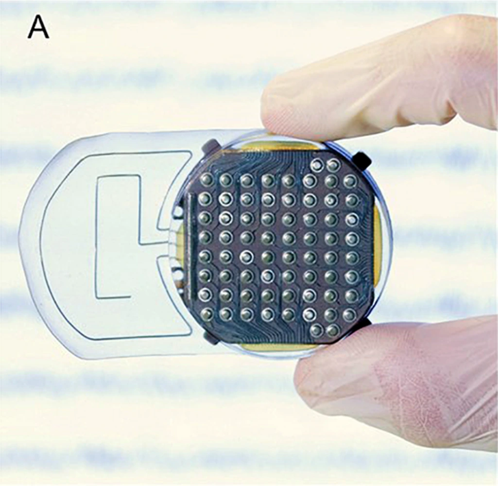
View closer → WIMAGINE implant viewed from its cortical-contact side, held between gloved fingers.
Hardware from the catalog’s cited 2019 preclinical study.
Image credit & source Sauter-Starace et al., Frontiers in Neuroscience (2019), Fig. 1A · Frontiers · Figure 1A · CC BY 4.0 .
Photographic panel cropped from the original figure; resized and re-encoded. Original scale bars and annotations retained.
Photograph
WIMAGINE (CEA-Clinatec) epidural wireless ECoG implant Open full-resolution image (1025 × 1000) →
WIMAGINE implant viewed from its cortical-contact side, held between gloved fingers.
Sauter-Starace et al., Frontiers in Neuroscience (2019), Fig. 1A · Source · CC BY 4.0 · Photographic panel cropped from the original figure; resized and re-encoded. Original scale bars and annotations retained.
Code L3?
Reviewed 2026-02-06
Interface ecog
Evidence stage human Independent, source-linked catalog sheet. Not a manufacturer-issued datasheet, regulatory decision or instructions for clinical use. Human evidence does not establish approval. Source-specific restrictions, conflicts and missing specifications are retained below.
Measured recordings Identity Geometry and architecture Electrode and channel physics Tissue interface and bioresponse System architecture Performance envelope Clinical and preclinical evidence Engineering tradeoffs References WIMAGINE (CEA-Clinatec) epidural wireless ECoG implant
The tables use the same field framework as the other implant-device sheets. Values belong to the named study or configuration. A blank cell means the reviewed sources do not establish a value, not that the device lacks that property.
The Clinatec exoskeleton trial (NCT02550522) covers the registry record and the published exoskeleton results.
Identity
Field Value and source scope Device WIMAGINE Manufacturer CEA-Clinatec, France; Sauter-Starace 2019 and 2015 development report Interface class Fully implanted wireless epidural ECoG recorder Origin CEA development; primary design published before the reviewed 2019 sheep study First demonstrated 2013 device conference report cited by the development literature; not a first implantation date First human implant June 2017 and November 2019 for the two patients in the 2021 stability study Species studied Sheep in 2019; humans in 2021 Regulatory status Research clinical protocol; conformity testing is not a claim of general market authorization Function Recording for motor BCI Target tissue Epidural surface above sensorimotor cortex
Geometry and architecture
Field Value and source scope Interface type Non-penetrating epidural ECoG Array layout 64 electrodes fixed beneath titanium cranial housing, 2019 Methods Electrode count 64 recording electrodes per implant, 2019 Methods Pitch 4 mm lateral and 4.5 mm anteroposterior, 2019 Methods Electrode lengths Not applicable; surface contacts Shank width and thickness Not applicable; implant fits a 50 mm craniotomy with 90 mm upper-surface curvature, 2019 Methods Tip and exposed site geometry 2.3 mm diameter contacts, 2019 Methods Contact coating Platinum-iridium 90/10 contact material, 2019 Methods Insulation Insertion method 50 mm craniotomy; contacts above intact dura, implant replaces removed bone, 2019 Methods Anchoring and fixation Four titanium wings protect against pressure or shocks, 2019 Methods; fixation details beyond these not reported
Electrode and channel physics
Field Value and source scope Exposed site area Electrode material Platinum-iridium 90/10, 2019 Methods Impedance (with measurement frequency) Noise floor or SNR 2015 development abstract: less than 0.7 µV RMS input-referred noise over 0.5-300 Hz; not a chronic in-vivo SNR Recording modality ECoG population field potentials Sampling rate 976 Hz, 12-bit ADC in 2019 sheep protocol; contacts recorded in successive 16-contact phases Stimulation capability Recording device; no therapeutic stimulation capability established Charge injection limit Not applicable to the reviewed recording use Reference and ground Reference electrodes shown in 2019 Figure 9; electrical topology not reported in this audit
Tissue interface and bioresponse
Field Value and source scope Target tissue Dura over sensorimotor cortex Insertion trauma and BBB disruption Epidural placement without cortical penetration; quantitative BBB disruption not reported Vascular disruption risk Micromotion sensitivity Gliosis and encapsulation 2019 sheep histology: increased GFAP reactivity in glia limitans and layer I under implant; do not describe as no gliosis Neuron loss near sites Foreign-body response mitigation Typical failure modes General failure rates not reported; 2021 measures signal stability, not lifetime reliability
System architecture
Field Value and source scope Onboard electronics Amplification and digitization beneath titanium housing, 2019/2015 descriptions Data path Wireless implant to external base station and laptop Telemetry bandwidth About 250 kb/s in 2-FSK mode, 2019 sheep protocol; MICS band 402-405 MHz Sampling rate 976 Hz in 2019 sheep protocol; 16 contacts at a time Power Remote inductive power at 13.56 MHz, 2019 Methods; not a rechargeable implanted battery claim Thermal management Packaging and hermeticity Hermetic titanium housing in 2015 development abstract MRI compatibility Surgical complexity 50 mm cranial opening and epidural module placement, 2019 Methods Output connectors Wireless base station to computer; no percutaneous electrode connector in described system
Field Value and source scope Acute yield Chronic yield No single yield percentage; two sheep retained relatively stable ECoG and SSEP over 10 months, 2019 Stability over time 2021 two-patient study: limited decline in RMS, band power and SNR; effective bandwidth and spectral edge frequency stable Longevity 32 and 14 months observed in two patients, 2021; not a maximum service life Revision and explant experience Postmortem explant and histology in two sheep, 2019; human revision rate not reported Adverse events Human adverse-event rate not reported in reviewed stability abstract; no generic safety claim Notable demonstrations Task-related motor imagery discrimination maintained beyond two years in 2021 study
Clinical and preclinical evidence
Field Value and source scope Human subjects Two bilaterally implanted quadriplegic patients, 2021 stability study Preclinical cohort Two sheep, 2019 study Follow-up duration Indications Motor BCI for paralysis research Trials and registries NCT02550522 is retained as the linked protocol record; current recruitment status not asserted Primary outcomes Longitudinal ECoG stability, effective bandwidth and evoked responses Key limitations Small cohorts, species-specific skull/contact geometry, 16-contact phased acquisition in sheep protocol
Engineering tradeoffs
Field Value and source scope Strengths Wireless fully implanted epidural acquisition with chronic human signal evidence Limitations Population recording, not single-unit access; histological reaction still documented in sheep Scaling constraints Wireless throughput, power and cranial footprint; no quantitative maximum channel count established beyond reviewed configuration
References
Recorded interface · Recorded with the WIMAGINE implant. Task annotations are instructions, not decoded movements; anatomical labels do not supply contact coordinates.
Explore eight recorded channels during executed and imagined reaching instructions. The released ECoG is filtered to 1–250 Hz; amplitude is shown in source units because physical calibration is not established.
Loading measured excerpt…
Enable JavaScript to load the measured excerpt. Source links remain available. Open the complete Recording Library →