Devices

Catalog specification sheet - Peripheral nerve

Macro-Sieve Electrode

Physical interfaceOptical micrograph
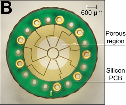
Circular macro-sieve electrode with a central porous gold-colored membrane, radial traces and eight peripheral connections; original 600-micrometre scale preserved.

Optical micrograph of the fabricated macro-sieve electrode and bonded micro-PCB; 2016 configuration.

Exact MacEwan et al. 2016 macro-sieve electrode in assigned catalog record. Panel crop, not a reconstruction. Include 600µm scale and region labels. License version to root-check through the article's actual CC BY link (4.0 expected).

Image credit & source

MacEwan, Zellmer, Wheeler, Burton & Moran, Frontiers in Neuroscience (2016), Figure 1B. DOI: 10.3389/fnins.2016.00557 · Frontiers · original figure · CC BY 4.0.

Photographic panel cropped from the original figure; resized and re-encoded. Original scale bars and annotations retained.

Optical micrograph

Macro-Sieve Electrode

Circular macro-sieve electrode with a central porous gold-colored membrane, radial traces and eight peripheral connections; original 600-micrometre scale preserved.

Open full-resolution image (503 × 421) →

Optical micrograph of the fabricated macro-sieve electrode and bonded micro-PCB; 2016 configuration.

MacEwan, Zellmer, Wheeler, Burton & Moran, Frontiers in Neuroscience (2016), Figure 1B. DOI: 10.3389/fnins.2016.00557 · Source · CC BY 4.0 · Photographic panel cropped from the original figure; resized and re-encoded. Original scale bars and annotations retained.

Code
?1R
Reviewed
2026-02-06
Interface
pni
Evidence stage
research

Independent, source-linked catalog sheet. Not a manufacturer-issued datasheet, regulatory decision or instructions for clinical use. Human evidence does not establish approval. Source-specific restrictions, conflicts and missing specifications are retained below.

Macro-sieve regenerative electrode with transit zones (MacEwan et al., 2016)

All rows follow the shared implant-device template. Measurements belong to the named study or configuration. A blank cell means the reviewed sources do not establish a value. Proposed use, terminal assays and continuously functioning implants are not treated as equivalent.

Identity

FieldValue and source scope
DeviceMacEwan 2016 macro-sieve electrode (MSE)
ManufacturerWashington University research fabrication, MacEwan 2016
Interface classRegenerative peripheral nerve sieve
OriginMacEwan, Zellmer, Wheeler, Burton and Moran team
First demonstrated2016 reviewed report; earliest prototype date not established
First human implantReviewed study uses rats
Species studiedAdult male Lewis rats
Regulatory statusPreclinical animal research
FunctionStimulation of regenerated sciatic nerve axons
Target tissueRegenerated sciatic nerve through sieve transit zones

Geometry and architecture

FieldValue and source scope
Interface typeRegenerative sieve between transected nerve ends
Array layout2 mm diameter porous region; nine transit zones around eight electrode sites
Electrode countEight Pt-Ir sites; four curved around central zone, four straight on alternating spokes
PitchNo uniform pitch reported; central transit hole about 600 µm diameter and each zone about 0.285 mm²
Electrode lengthsNot applicable to shanks; two 3 mm silicone conduit segments attached to sieve
Shank width and thicknessPolyimide wafer thickness not reported in audited article; assembly elastomer encapsulation 1-2 mm is not wafer thickness
Tip and exposed site geometryCurved central sites about 33,000 µm²; straight peripheral sites about 24,000 µm²
Contact coatingPt-Ir metallized/platinized sites as described by article; terminology preserved
InsulationPolyimide base, medical epoxy at connections and elastomer encapsulation; silicone nerve conduits
Insertion methodTransect sciatic nerve, secure stumps in conduit assembly with about 4 mm gap
Anchoring and fixationNerve stumps secured inside conduits; connectors placed subcutaneously for terminal re-exposure

Electrode and channel physics

FieldValue and source scope
Exposed site areaAbout 33,000 µm² central curved and 24,000 µm² peripheral straight sites, Results
Electrode materialPt-Ir/platinized active sites with gold traces on polyimide, Figures/Methods
Impedance (with measurement frequency)At 1 kHz before implantation: 2.3-6.3 kΩ, mean 4.2 ± 0.9 kΩ; not chronic impedance
Noise floor or SNR
Recording modalityElectrical stimulation through MSE; CNAP/EMG assay recordings use separate electrodes, not chronic MSE neural recording
Sampling rate40 kHz CNAP and 10 kHz EMG assay acquisition; not MSE onboard sampling specifications
Stimulation capabilityMonopolar individual-site and simultaneous-site stimulation evoked reinnervated muscle force at three months
Charge injection limit
Reference and groundMonopolar assay configuration; exact assembly reference/ground pin map not reported

Tissue interface and bioresponse

FieldValue and source scope
Target tissueRegenerating peripheral sciatic nerve
Insertion trauma and BBB disruptionBBB: not applicable; intentional sciatic nerve transection
Vascular disruption riskMicrovasculature seen crossing MSE; quantified vascular injury rate not reported
Micromotion sensitivityMicro-PCB increases rigidity, Results; quantified micromotion not reported
Gliosis and encapsulationMyelinated/unmyelinated fibers through transit zones; quantitative fibrosis burden not reported
Neuron loss near sites
Foreign-body response mitigationLarge transit zones; GDNF tested versus saline and conduit controls, not universal coating efficacy
Typical failure modesNo assembly damage/failure observed through three months; longer-term failure rate not reported

System architecture

FieldValue and source scope
Onboard electronicsPassive electrode on micro-PCB; external stimulator for terminal evaluation
Data pathGold traces to micro-PCB/microwires, subcutaneous Omnetics connector re-exposed at terminal experiment
Telemetry bandwidthNot applicable; wired assay
Sampling rateExternal assay-specific, no onboard ADC
PowerExternal test equipment
Thermal management
Packaging and hermeticityEpoxy EpoTek 730 protects connections; elastomer MDX4-4210 encapsulation; no hermetic certification
MRI compatibility
Surgical complexityNerve transection, conduit assembly, lead routing and subcutaneous connector pocket
Output connectorsFemale Omnetics 20-pin connector, Methods

Performance envelope

FieldValue and source scope
Acute yieldAll tested metallized sites functional at implantation, Results; tested-site denominator not reported
Chronic yieldAssembly integrity maintained in all implanted animals through three months; not channel-by-channel chronic recording yield
Stability over timeThree-month terminal stimulation evidence; longitudinal recording stability not reported
LongevityThree months observed, not maximum lifetime
Revision and explant experienceTerminal explant/histology, no clinical revision cohort
Adverse eventsNo assembly damage/failure reported in study; nerve injury inherent to model, not a general safety claim
Notable demonstrationsIndividual MSE sites selectively recruited reinnervated muscles; all-site stimulation evoked force comparable to epineurial stimulation

Clinical and preclinical evidence

FieldValue and source scope
Human subjectsNot applicable to reviewed preclinical study
Preclinical cohort40 rats randomized to five groups of eight; two MSE groups of eight, two conduit groups and unoperated controls
Follow-up durationThree months, terminal physiology; gait tracked at one, two and three months
IndicationsPreclinical regenerative peripheral interface
Trials and registriesAnimal research; human registry not applicable
Primary outcomesHistomorphometry, CNAP, EMG, muscle force and behavior
Key limitationsRat regenerative capacity and short follow-up limit human inference; terminal assay not continuous implanted recording

Engineering tradeoffs

FieldValue and source scope
StrengthsLarge transit zones with regenerated axons and selective stimulation at three months
LimitationsTransection required; PCB stiffens thin sieve; no long-term human or recording evidence
Scaling constraintsEight large sites access sectors rather than isolate each axon; density versus transit-area tradeoff

References