Science neuronal-embedded electronic biohybrid interface (architecture concept)
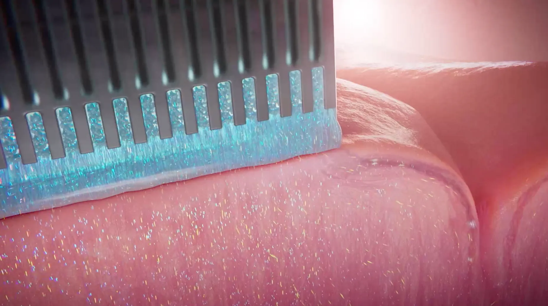
Science electronic biohybrid architecture concept: device-associated neurons and their brain connections. Company illustration, without a measured form factor.
Company architecture concept illustration; not a physical hardware photograph or a measured as-built blueprint. The separate passive microwell scaffold is not substituted.
Scale: Concept illustration without a scale bar.
Schematic rendering only; dimensions and electrode/site geometry are not established by this illustration.
Image credit & source
Science Corporation, Biohybrid neural interfaces (23 November 2024) · Science Corporation · architecture illustration · Copyright © Science Corporation; reserved-rights reference.
Company source illustration retained intact; no generative changes.
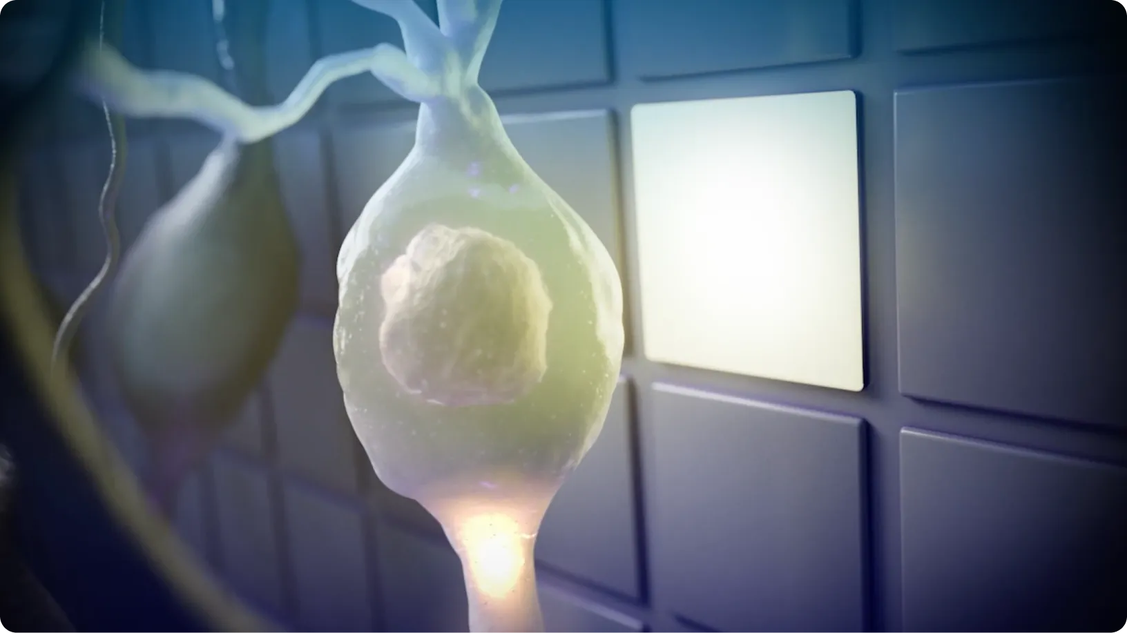
MicroLED-to-neuron interaction in the proposed biohybrid electronic architecture. Concept rendering; no calibrated dimensions.
Company architecture concept illustration; not a physical hardware photograph or a measured as-built blueprint. The separate passive microwell scaffold is not substituted.
Scale: Concept illustration without a scale bar.
Schematic rendering only; dimensions and electrode/site geometry are not established by this illustration.
Image credit & source
Science Corporation, Biohybrid neural interfaces (23 November 2024) · Science Corporation · microLED illustration · Copyright © Science Corporation; reserved-rights reference.
Company source illustration retained intact; no generative changes.
- Code
- ?0R
- Reviewed
- 2026-02-06
- Interface
- other
- Evidence stage
- theoretical
Independent, source-linked catalog sheet. Not a manufacturer-issued datasheet, regulatory decision or instructions for clinical use. Human evidence does not establish approval. Source-specific restrictions, conflicts and missing specifications are retained below.
Science neuronal-embedded electronic biohybrid interface (architecture concept)
All rows follow the shared implant-device template. Measurements belong to the named study or configuration. A blank cell means the reviewed sources do not establish a value. Proposed architectures, optical behavior and validated electronic recording systems are kept separate.
Identity
| Field | Value and source scope |
|---|---|
| Device | Science neuronal-embedded electronic biohybrid probe architecture concept; penetrating form factor not established |
| Manufacturer | Science Corporation, company description November 2024 |
| Interface class | Conceptual biohybrid neural interface; deployment geometry not reported |
| Origin | Science Corporation biohybrid program led by Alan Mardinly and Yifan Kong |
| First demonstrated | Company architecture description 2024; complete electronic system demonstration not established |
| First human implant | |
| Species studied | No cohort for this complete electronic architecture given; separate surface microwell study uses mice |
| Regulatory status | Company describes low technology readiness; clinical authorization not reported |
| Function | Proposed electrical interaction with device-associated neurons and optical activation through microLEDs; not a validated bidirectional system |
| Target tissue | Brain via graft neurites; precise surgical target/placement not reported |
Geometry and architecture
| Field | Value and source scope |
|---|---|
| Interface type | Neurons embedded in electronics with neurites growing into host tissue; penetrating shanks not established |
| Array layout | |
| Electrode count | Company million-neuron/billion-synapse examples are scaling ambitions, not device/contact counts |
| Pitch | |
| Electrode lengths | |
| Shank width and thickness | No dimensions transferred from separate 5 x 5 mm microwell scaffold |
| Tip and exposed site geometry | |
| Contact coating | |
| Insulation | |
| Insertion method | Source does not establish a penetrating probe or insertion procedure |
| Anchoring and fixation |
Electrode and channel physics
| Field | Value and source scope |
|---|---|
| Exposed site area | |
| Electrode material | |
| Impedance (with measurement frequency) | |
| Noise floor or SNR | Company calls SNR great without a numerical measurement for this architecture; not treated as a measured spec |
| Recording modality | Electrical interaction with graft cells proposed; validated on-device recording yield/modality not reported |
| Sampling rate | |
| Stimulation capability | Company illustration shows microLED activating nearby neuron; wavelength, power and complete-system validation not reported |
| Charge injection limit | |
| Reference and ground |
Tissue interface and bioresponse
| Field | Value and source scope |
|---|---|
| Target tissue | Device-associated graft neurons and their axons/dendrites in brain |
| Insertion trauma and BBB disruption | Quantitative trauma and BBB effects not reported for this architecture; not presumed noninvasive |
| Vascular disruption risk | |
| Micromotion sensitivity | |
| Gliosis and encapsulation | |
| Neuron loss near sites | |
| Foreign-body response mitigation | Company identifies hypoimmunogenic allogeneic neurons as a development need, not a demonstrated clinical solution |
| Typical failure modes | Company identifies graft survival under hypoxia, glycemic shock and host immunity as engineering challenges; failure rates not published here |
System architecture
| Field | Value and source scope |
|---|---|
| Onboard electronics | Electronics with embedded neurons and illustrated microLED; circuit configuration not reported |
| Data path | |
| Telemetry bandwidth | |
| Sampling rate | |
| Power | Company argues strong cell signals could reduce amplifier power; measured system power not reported |
| Thermal management | |
| Packaging and hermeticity | |
| MRI compatibility | |
| Surgical complexity | Implant procedure not reported; no geometry inferred from the word probe |
| Output connectors |
Performance envelope
| Field | Value and source scope |
|---|---|
| Acute yield | |
| Chronic yield | |
| Stability over time | Complete electronic architecture performance not reported; mouse microwell survival not transferred |
| Longevity | |
| Revision and explant experience | |
| Adverse events | No safety cohort for this architecture; surface microwell explant injury belongs to that separate study |
| Notable demonstrations | Company links a separate passive cortical microwell/optical mouse proof of concept; not proof of this electronic probe system |
Clinical and preclinical evidence
| Field | Value and source scope |
|---|---|
| Human subjects | |
| Preclinical cohort | Not given for this architecture; Brown mouse microwell cohort is separate |
| Follow-up duration | |
| Indications | Long-term high-bandwidth brain interface ambition, not validated clinical indication |
| Trials and registries | |
| Primary outcomes | No quantitative complete-system outcomes published in reviewed company description |
| Key limitations | Architecture-level company source; no pinned form factor, contact map, chronic electronics or clinical evidence. Surface scaffold findings must not be relabeled as probe results |
Engineering tradeoffs
| Field | Value and source scope |
|---|---|
| Strengths | Proposed biological coupling through neurons and synapses; theoretical scaling path |
| Limitations | Cell production, immune compatibility, survival and electronic packaging remain unresolved in public specification |
| Scaling constraints | Million-neuron and billion-synapse company examples are not measured throughput or channel count |
References
- Science Corporation, Biohybrid neural interfaces, November 2024. Company architecture description and illustration; future-scale examples are not measured specifications.
- Brown et al. Separate surface microwell study and full manuscript. Context only; evidence belongs to its surface scaffold, not this unspecified electronic probe architecture.