FINE (Flat Interface Nerve Electrode)
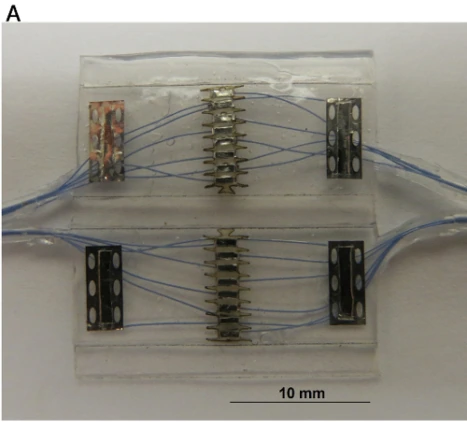
Fabricated 16-contact FINE in its open position, showing the tissue-facing contacts and reference strips. 10 mm scale retained.
2016 high-contact-density academic fabrication example; not the separate Ardiem human sensory cuff.
Scale: 10 mm scale bar retained.
Open, flat fabrication configuration; closed-cuff aperture must be obtained from the paper specifications rather than inferred from this open photograph.
Image credit & source
Dweiri et al., Journal of Visualized Experiments (2016), Fig. 3A · Dweiri 2016 · Fig. 3A · Copyright reserved; educational reference use requested by site owner.
Photographic panel mechanically cropped from the source; resized and encoded as WebP. Source annotations and scale bars retained; no generative changes.
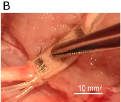
FINE around the canine sciatic nerve, showing the longitudinally flexible cuff body; 10 mm scale retained.
2016 high-contact-density academic fabrication example; not the separate Ardiem human sensory cuff.
Scale: 10 mm source scale bar retained.
Surgical orientation photograph; tissue and cuff may be deformed by handling.
Image credit & source
Dweiri et al. (2016), Fig. 3B · Dweiri 2016 · Fig. 3B · Copyright reserved; educational reference use requested by site owner.
Photographic panel mechanically cropped from the source; resized and encoded as WebP. Source annotations and scale bars retained; no generative changes.
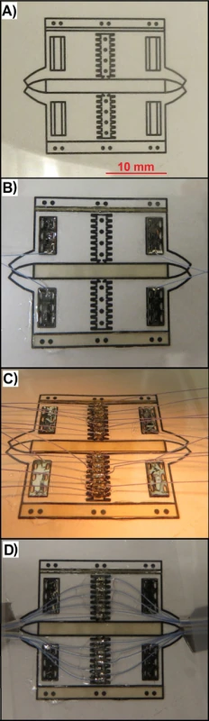
FINE fabrication sequence: tissue-facing contact layout and lead routing before final encapsulation.
2016 high-contact-density academic fabrication example; not the separate Ardiem human sensory cuff.
Scale: No calibrated scale bar in this process figure.
Intermediate fabrication views; not an additional clinical cuff version.
Image credit & source
Dweiri et al. (2016), Fig. 2 · Dweiri 2016 · Fig. 2 · Copyright reserved; educational reference use requested by site owner.
Complete source figure retained; resized and encoded as WebP. No generative changes.
- Code
- ?3R
- Reviewed
- 2026-02-06
- Interface
- pni
- Evidence stage
- human
Independent, source-linked catalog sheet. Not a manufacturer-issued datasheet, regulatory decision or instructions for clinical use. Human evidence does not establish approval. Source-specific restrictions, conflicts and missing specifications are retained below.
FINE (Flat Interface Nerve Electrode)
Every field follows the shared implant-device template. Values belong to a named configuration or study; not reported means the reviewed sources do not establish the value. Family-wide and deployment-specific evidence are kept separate.
Identity
| Field | Value and source scope |
|---|---|
| Device | FINE family; human sensory cuffs and 2016 high-contact-density fabrication kept distinct |
| Manufacturer | Tan 2015 human components: Ardiem Medical. Dweiri 2016 fabrication is an academic protocol, not the same manufactured cuff |
| Interface class | Extraneural flat-interface peripheral nerve cuff |
| Origin | Tyler and Durand design cited in Dweiri 2016 |
| First demonstrated | 2002 foundational selective-stimulation paper cited in Dweiri 2016; earliest prototype date not reported |
| First human implant | Tan 2015 reports first chronic human FINE study; earliest exact implantation date not established in this audit |
| Species studied | Human in Tan 2015; species of 2016 chronic fabrication measurement not pinned here |
| Regulatory status | Tan 2015 research under FDA IDE; not general market approval |
| Function | Selective stimulation and electroneurogram recording, configuration-specific |
| Target tissue | Peripheral nerve surface |
Geometry and architecture
| Field | Value and source scope |
|---|---|
| Interface type | Non-penetrating cuff with flat cross-section |
| Array layout | Tan 2015 eight-contact FINEs; Dweiri 2016 contact arrays framed with PEEK and paired outer references |
| Electrode count | Eight per FINE in Tan 2015; 2016 high-density design count configuration dependent, not inferred from images |
| Pitch | 2016 middle-contact spacing 0.51 ± 0.04 mm, n=70; not a clinical Tan 2015 pitch |
| Electrode lengths | Not applicable to shanks; axial cuff length configuration dependent |
| Shank width and thickness | 2016 PEEK contact frame 125 µm thick, not total cuff thickness. Tan 2015 lumen 10 x 1.0 or 10 x 1.5 mm, depending on nerve/subject |
| Tip and exposed site geometry | 2016 laser-cut strip width follows frame guide channel; numerical exposed active area not reported |
| Contact coating | Platinum/10% iridium strips in 2016 protocol; not automatically assigned to Tan 2015 clinical cuffs |
| Insulation | 2016 silicone cuff surrounding 125 µm PEEK contact frame; clinical cuff stack not established here |
| Insertion method | Surgical nerve exposure, placement around nerve without penetrating epineurium |
| Anchoring and fixation | 2016 protocol sutures two free edges together; one human ulnar FINE opened after surgery in Tan 2015 |
Electrode and channel physics
| Field | Value and source scope |
|---|---|
| Exposed site area | |
| Electrode material | Platinum/10% iridium contacts, Dweiri 2016; human clinical contact material not reported in audited Tan Methods |
| Impedance (with measurement frequency) | Dweiri 2016: 2.55 ± 0.25 kΩ at 7.5 months, measurement frequency not reported in audited abstract. Tan 2015 pairwise pulse-derived values about 2.66-3.12 kΩ, measured with 0.3 mA/50 µs pulses at 20/100 Hz; not one 1 kHz rating |
| Noise floor or SNR | 2016 protocol: chronic recording SNR 5.10 ± 0.81 dB at 7.5 months; not a device-wide noise floor |
| Recording modality | Peripheral electroneurogram in 2016 fabrication report; Tan 2015 concerns stimulation |
| Sampling rate | Configuration dependent; 2016 external preamplifier bandwidth 700 Hz-7 kHz and gain 2,000 do not establish sample rate |
| Stimulation capability | Selective sensory stimulation in Tan 2015; 2016 protocol supports recording/stimulation configurations |
| Charge injection limit | Not given as material limit; perceptual thresholds are not charge-injection limits |
| Reference and ground | 2016 protocol: four outer reference contacts and middle arrays, positioned symmetrically for interference rejection; clinical topology not equated |
Tissue interface and bioresponse
| Field | Value and source scope |
|---|---|
| Target tissue | Nerve surface, outside epineurium |
| Insertion trauma and BBB disruption | BBB: not applicable; extraneural cuff placement. Quantitative injury not reported in audited human study |
| Vascular disruption risk | Compression/fit is a design concern; no quantitative human vascular injury rate |
| Micromotion sensitivity | 2016 design balances transverse rigidity and longitudinal flexibility; quantitative motion sensitivity not reported |
| Gliosis and encapsulation | CNS gliosis not applicable; Tan 2015 discusses encapsulation as possible mechanism, not quantitative human histology |
| Neuron loss near sites | |
| Foreign-body response mitigation | 2016 design expands cross-sectional area by 25% at 67 mmHg bench pressure; not an approved safe nerve-compression limit |
| Typical failure modes | Tan 2015: ulnar FINE opened in subject 2, losing intimate nerve contact; other contacts remained available |
System architecture
| Field | Value and source scope |
|---|---|
| Onboard electronics | None in cuff; external acquisition/stimulation electronics in audited configurations |
| Data path | Tan 2015 cuff leads through spring-sleeve connectors to percutaneous leads and external UECU |
| Telemetry bandwidth | Not applicable to passive wired cuff |
| Sampling rate | Configuration dependent; external acquisition |
| Power | External stimulation/acquisition system in audited configurations |
| Thermal management | |
| Packaging and hermeticity | 2016 silicone/PEEK passive cuff; Tan 2015 uses percutaneous leads, not a fully sealed implant |
| MRI compatibility | |
| Surgical complexity | Peripheral nerve exposure with careful lumen selection and closure |
| Output connectors | Tan 2015 spring-sleeve to open-helix percutaneous leads; 2016 connector model not reported |
Performance envelope
| Field | Value and source scope |
|---|---|
| Acute yield | Tan 2015 subject 2: initially 10/16 available contacts evoked sensation; excludes opened ulnar cuff. Not a family-wide yield |
| Chronic yield | Tan 2015 subject 2: 16/16 available contacts by week 27. Subject 1 total 19/20 includes four spiral contacts, so not assigned wholly to FINE |
| Stability over time | Tan 2015 threshold/impedance/percept evidence over 1-2 years in mixed cuff study; configurations distinguished |
| Longevity | 2016 recording cuff observed at 7.5 months; human sensory study 1-2 years, not maximum lifetime |
| Revision and explant experience | Opened human ulnar cuff documented; revision/explant details not reported |
| Adverse events | Tan 2015: no infection of percutaneous leads or implanted components in study period; one FINE opened |
| Notable demonstrations | Characteristic sensory percept fields in human amputees; 2016 reports chronic neural recording |
Clinical and preclinical evidence
| Field | Value and source scope |
|---|---|
| Human subjects | Tan 2015: two amputees with FINEs; subject 1 also has a spiral radial cuff. Do not attribute mixed-system totals to FINE alone |
| Preclinical cohort | 2016 chronic recording example; cohort size/species not reported in audited abstract |
| Follow-up duration | Tan 2015: 1-2 years; 2016 chronic recording example: 7.5 months |
| Indications | Experimental sensory restoration after upper-limb loss and peripheral nerve recording |
| Trials and registries | Tan 2015 FDA IDE; registry identifier not reported |
| Primary outcomes | Percept selectivity, thresholds, pulse-derived impedance, chronic recording SNR |
| Key limitations | FINE is a family with different builds; tiny human cohort, mixed cuff types, opened cuff and nonuniform measurement methods |
Engineering tradeoffs
| Field | Value and source scope |
|---|---|
| Strengths | Multiple surface contacts with human selective sensory evidence; high-density fabrication and chronic recording reported separately |
| Limitations | Fit and closure can fail; extraneural population recording is not single-axon access |
| Scaling constraints | Transverse shaping versus longitudinal flexibility, lead count and fabrication precision; no universal scaling maximum |
References
- Dweiri et al. 2016. Fabrication of High Contact-Density, Flat-Interface Nerve Electrodes for Recording and Stimulation Applications. Protocol, not a universal clinical specification.
- Tan et al. 2015. Stability and selectivity of a chronic, multi-contact cuff electrode for sensory stimulation in human amputees. Human FINE and spiral configurations separated.
Models and reference visualizations
FINE · 2013 eight-contact lumen reference
Electrode-bearing lumen; simplified silicone shell · dimensions in millimeters
Loading 3D viewer…
Documented measurements
- 10 × 1.5 × 7 mm selected lumen
- Eight 0.5 mm apertures, approximately 2 mm pitch and opposing half-pitch stagger
Reconstructed or assumed
- 0.3 mm wall; photo-informed rounded lumen and exterior
- Centered transverse row origins and axial midpoint
Not modeled
- Contact recess, snap closure, strain relief and leads
- Nerve reshaping and electrical response
Model fidelity. Selected 10 × 1.5 × 7 mm luminal variant from Schiefer et al. (2013), with four inward-facing 0.5 mm apertures on each opposing wall. Approximately 2 mm center spacing and half-pitch staggering are reported. Absolute X offsets are centered for display and axial centers are placed at the cuff midpoint; those origins were not specified. The 0.3 mm wall thickness and rounded corners are display assumptions, not measured silicone geometry. The 2013 source photograph shows rounded lumen ends and a soft outer silhouette: the displayed 0.75 mm inner radius and 0.3 mm outer radius preserve the documented 10 × 1.5 mm lumen envelope and contact locations. Pale silicone and silver Pt/Ir reflect the documented materials; optical finish is illustrative. Recess depth, closure, lead exits and strain relief are omitted. Contact area describes ideal aperture area, not electrochemical surface area. Not the 2016 high-density PEEK-frame FINE. No nerve reshaping, pressure or stimulation is calculated.
Color, transparency and surface finish approximate the references. Use Highlight contacts to inspect electrode locations.
Contact meshes use a 10 nm display separation to prevent surface flicker in 3D exports. Geometry JSON retains the nominal measured surfaces.
Sources & reference images
Dimensions come from the linked specifications; images guide shape only. Reference images remain on their owners’ websites.
JSON includes physical site coordinates, unknown channel mappings and approximation notes. GLB uses meters. These models do not predict recording or stimulation performance.