TU Delft transfer-free multilayer graphene MEA (DBI2 TS4)
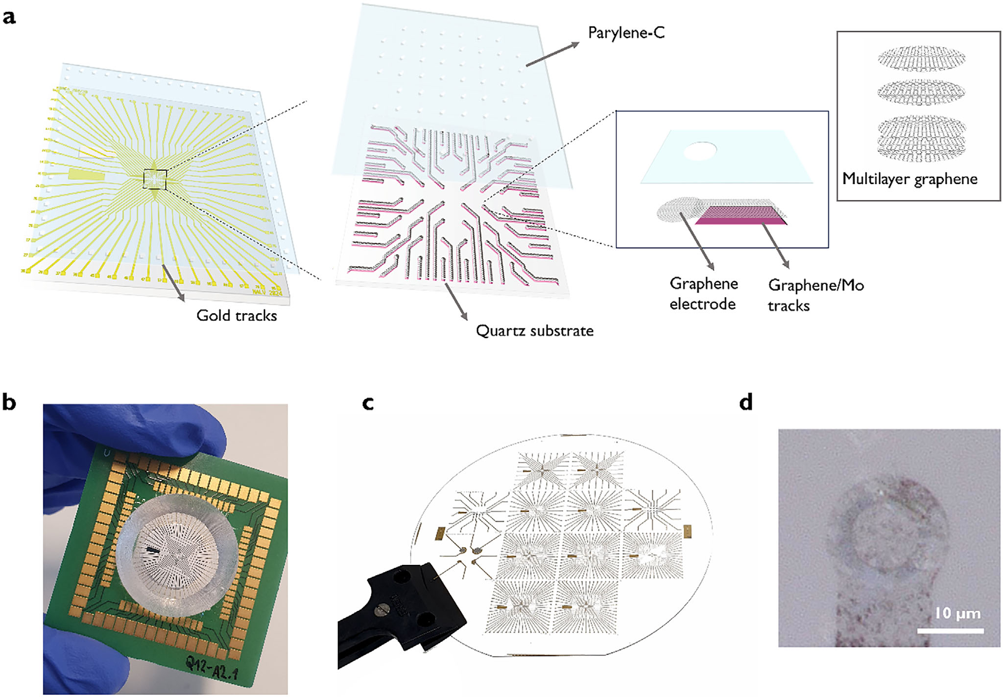
Complete Figure 1: rigid fused-silica bench MEA and electrode close-up, tested on brain slices; this device is not an implant.
Scale: Original 10 µm scale bar retained in panel d. The other panels use independent scales.
In vitro brain-slice MEA on rigid fused silica; no in vivo implantation is demonstrated.
Image credit & source
Alvarez de Eulate et al., Advanced Science (2026), Figure 1 · Alvarez de Eulate et al., Advanced Science (2026), Figure 1 · CC BY 4.0.
Complete original figure extracted from the final published PDF hosted by TU Delft. No crop, resizing, annotation or image enhancement.
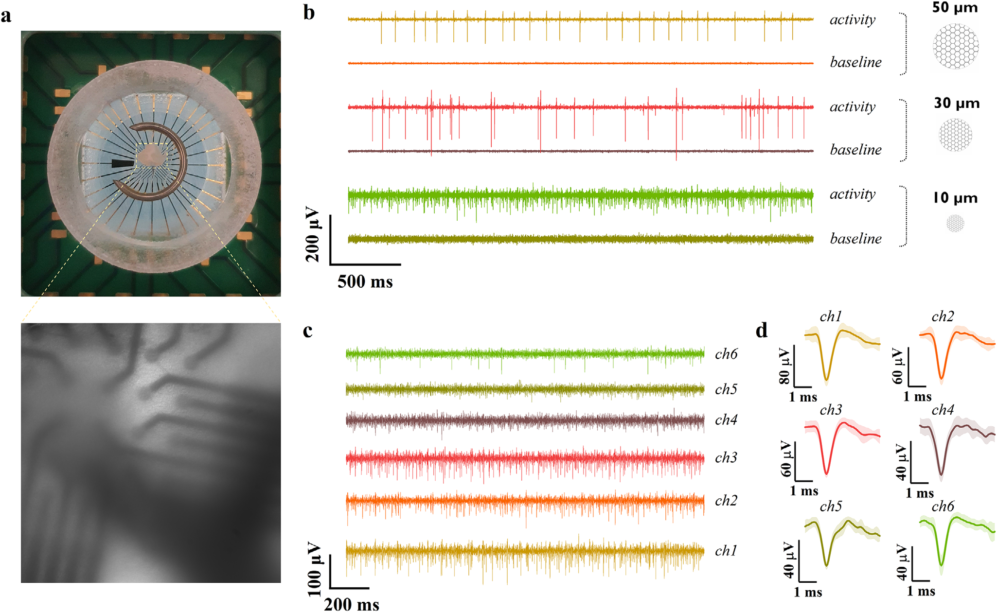
Complete Figure 3 shows ex vivo brain-slice placement and recording data; it does not show an implanted device.
Scale: Original panel scales retained. The recording chamber is a bench apparatus.
Ex vivo brain-slice interface; retained in the existing catalog with an explicit non-implant caveat.
Image credit & source
Alvarez de Eulate et al., Advanced Science (2026), Figure 3 · Alvarez de Eulate et al., Advanced Science (2026), Figure 3 · CC BY 4.0.
Complete original figure extracted from the final published PDF hosted by TU Delft. No crop, resizing, annotation or image enhancement.
- Code
- L1R
- Reviewed
- 2026-10-14
- Interface
- other
- Evidence stage
- research
Independent, source-linked catalog sheet. Not a manufacturer-issued datasheet, regulatory decision or instructions for clinical use. Human evidence does not establish approval. Source-specific restrictions, conflicts and missing specifications are retained below.
TU Delft transfer-free multilayer graphene MEA (DBI2 TS4)
A transparent microelectrode array whose recording sites are multilayer graphene grown in place on the substrate, instead of transferred from a pre-grown sheet. It is an in vitro research tool for combining electrical recording with optical imaging, tested on brain slices. It is not an implant.
Identity
| Field | Value and source scope |
|---|---|
| Device | Transfer-free multilayer graphene MEA on transparent fused silica [1] |
| Origin | Delft University of Technology (Department of Microelectronics), with Fraunhofer IZM Berlin and Erasmus MC Rotterdam listed among author affiliations [1] |
| Programme | Dutch Brain Interface Initiative, task TS4 (PI Vasiliki Giagka, TU Delft); the paper states it is part of the DBI2 project [1][3] |
| Radboud and Donders link | The DBI2 annual report says graphene electrode designs were developed in collaboration with the Battaglia lab at Radboud University [3]. No Radboud or Donders author appears on the paper’s author list [1] |
| Species | Mouse (acute cerebellar slices) [1] |
| Regulatory status | Research device, in vitro [1] |
Geometry and architecture
| Field | Value and source scope |
|---|---|
| Substrate | Transparent fused silica wafer; the 4 in wafer carried 12 MEAs of different designs [1] |
| Array layout | Three primary designs: 60 electrodes of 10 µm in an 8 x 8 grid at 100 µm pitch; 60 electrodes of 50 µm in an 8 x 8 grid at 200 µm pitch; 32 electrodes of 30 µm in a 6 x 6 grid at 100 µm pitch. A further design spans 10 to 500 µm diameters [1] |
| Electrode diameters | 10, 30 and 50 µm tested on slices [1] |
| Tracks and pads | Inner tracks inside the imaging region are a graphene and molybdenum bilayer; outer tracks and pads are Ti/Au [1] |
| Insulation | Parylene-C, 1 to 2 µm [1] |
| Interface | Custom PCB sized for Multichannel Systems 60-electrode MEA recording systems, also compatible with a TDT MZ60 interface [1] |
Materials and fabrication
| Field | Value and source scope |
|---|---|
| Conductor | Multilayer turbostratic graphene grown by CVD on a molybdenum catalyst layer on the fused silica, so no transfer step is needed [1] |
| Layer count | Estimated at roughly 20 to 25 layers from 47.5% transmittance at 550 nm, assuming about 2.3% absorption per layer; the authors call this a linear approximation [1] |
| Transparency | About 50% across the spectrum; 47.5% at 550 nm [1] |
Performance envelope
| Field | Value and source scope |
|---|---|
| Impedance | 5.68 ± 1.37 MΩ at 1 kHz for 10 µm electrodes (n = 42); other sizes in the paper’s supplementary table, not extracted here [1] |
| Noise | Baseline rms noise 2 to 7 µV [1] |
| Spike amplitude | Up to about 140 µV [1] |
| Signal to noise | Published article: 18.6 ± 2.7 dB (10 µm), 24.4 ± 6.2 dB (30 µm), 24.2 ± 4.3 dB (50 µm); abstract states up to about 25 dB. The 2025 preprint abstract states up to 30 to 40 dB [1][2] |
| Recording result | Spontaneous extracellular spiking from acute mouse cerebellar slices, which the authors attribute to Purkinje cells; the paper says signal quality was limited more by electrode-tissue coupling than by the MEA technology [1] |
| First claimed | The published abstract says it is the first demonstration that transparent graphene electrodes as small as 10 µm record extracellular spiking at these signal-to-noise ratios [1] |
| Annual-report status | Year 2 report: graphene MEAs with 30 µm electrodes on transparent substrates recorded spikes from brain slices; two-photon tests confirmed graphene sheet transparency [3] |
Limits
In vitro slice recordings only; no implantation, animal in vivo recording or chronic data. The DBI2 task TS4 aims at flexible, stretchable, freely floating active arrays; the device here is on rigid fused silica. The paper says a flexible-substrate version of the transfer-free process was reported earlier; that version is not catalogued here. Charge injection, long-term stability and biocompatibility testing are blank.
References
- Alvarez de Eulate N, Pavlou C, León González G, Camarena Pérez M, Holzapfel L, Gao Z, Vollebregt S, Giagka V. Transparent transfer-free ultrasmall multilayer graphene microelectrodes enable high quality recordings in brain slices. Advanced Science 13(20):e17524 (2026), CC BY. DOI 10.1002/advs.202517524; full text PMC13067786.
- Preprint of the same work, bioRxiv 2025, DOI 10.1101/2025.06.13.657566, listed on the DBI2 publications page.
- DBI2 Annual Report, Year 2, Work Package 3, task TS4.
- DBI2 project page: graphene-based neural interfaces (TU Delft postdoc project, September 2023 to August 2027, owner Christos Pavlou).