TIME (Transverse Intrafascicular Multichannel Electrode)
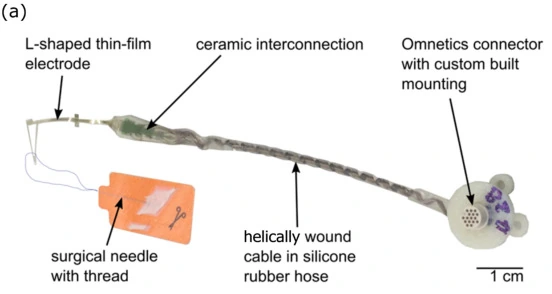
TIME-4H assembly with thin-film electrode and helical lead; 1 cm scale retained.
TIME-4H configuration (2021). The connector and interconnect are shown alongside the implantable thin-film electrode.
Image credit & source
Guiho et al., Sensors (2021), Fig. 2a · Sensors · Figure 2a · CC BY 4.0.
Photographic panel cropped from the original figure; resized and re-encoded. Original scale bars and annotations retained.
- Code
- ?3?
- Reviewed
- 2026-02-03
- Interface
- pni
- Evidence stage
- human
Independent, source-linked catalog sheet. Not a manufacturer-issued datasheet, regulatory decision or instructions for clinical use. Human evidence does not establish approval. Source-specific restrictions, conflicts and missing specifications are retained below.
TIME - Transverse Intrafascicular Multichannel Electrode
The tables use the same field framework as the other implant-device sheets. Values belong to the named study or configuration. A blank cell means the reviewed sources do not establish a value, not that the device lacks that property.
Identity
| Field | Value and source scope |
|---|---|
| Device | TIME; geometry below is TIME-4H in the 2021 STIMEP study |
| Manufacturer | TIME developed by Laboratory for Biomedical Microtechnology, IMTEK, University of Freiburg; 2021 Methods |
| Interface class | Transverse intrafascicular peripheral nerve electrode |
| Origin | Boretius et al. 2010 primary design report |
| First demonstrated | 2010 primary report, not a claim of earliest prototype date |
| First human implant | |
| Species studied | Rats in Boretius 2010 and STIMEP 2021; human amputee in Raspopovic 2014 |
| Regulatory status | Research device; 2021 design for human trials is not commercial approval |
| Function | Selective peripheral nerve stimulation; original 2010 report also characterizes recording transfer behavior |
| Target tissue | Peripheral nerve fascicles; sciatic nerve in reviewed rat experiments |
Geometry and architecture
| Field | Value and source scope |
|---|---|
| Interface type | Penetrating transverse intrafascicular thin film |
| Array layout | TIME-4H folded U-to-L thin film: seven stimulation sites and one ground on each side, 2021 Methods |
| Electrode count | 14 independent stimulation sites and two ground sites, TIME-4H 2021 |
| Pitch | 0.75 mm within a side, opposite side offset 0.375 mm, TIME-4H 2021 |
| Electrode lengths | |
| Shank width and thickness | |
| Tip and exposed site geometry | 80 µm diameter active openings; each ground comprises 109 connected 80 µm contacts, TIME-4H 2021 |
| Contact coating | Highly porous sputtered iridium oxide on active sites, TIME-4H 2021 |
| Insulation | Polyimide thin-film substrate and insulation; PEI insulated cable, silicone protection, 2021 Methods |
| Insertion method | Attached needle and suture sling thread electrode transversely through nerve, TIME-4H 2021 |
| Anchoring and fixation |
Electrode and channel physics
| Field | Value and source scope |
|---|---|
| Exposed site area | TIME-4H active-site diameter 80 µm; ground aggregate about 0.55 mm², 2021 Methods. Active area not separately reported |
| Electrode material | Platinum tracks, silicon carbide adhesion promoter, SIROF active contacts, TIME-4H 2021 |
| Impedance (with measurement frequency) | |
| Noise floor or SNR | |
| Recording modality | Original 2010 report characterizes recording transfer; 2021 study measures muscle CMAP with separate EMG electrodes, not TIME neural recording |
| Sampling rate | Configuration dependent; 20 kHz in 2021 is external EMG acquisition, not a TIME implant sampling spec |
| Stimulation capability | Selective stimulation demonstrated in rat sciatic nerve, 2010 and 2021 |
| Charge injection limit | |
| Reference and ground | Two ground sites, each formed from 109 interconnected openings, TIME-4H 2021 |
Tissue interface and bioresponse
| Field | Value and source scope |
|---|---|
| Target tissue | Peripheral nerve fascicles |
| Insertion trauma and BBB disruption | BBB: not applicable to peripheral nerve; quantitative nerve insertion injury not reported in these acute studies |
| Vascular disruption risk | |
| Micromotion sensitivity | |
| Gliosis and encapsulation | |
| Neuron loss near sites | |
| Foreign-body response mitigation | |
| Typical failure modes | Chronic failure rates not reported; 2010 report explicitly leaves chronic material-tissue behavior to future work |
System architecture
| Field | Value and source scope |
|---|---|
| Onboard electronics | Passive thin-film electrode connected to external stimulator, 2021 Methods |
| Data path | Wired, 10 cm cable with 16 helically wound MP35N wires, TIME-4H 2021 |
| Telemetry bandwidth | Not applicable to the passive wired electrode |
| Sampling rate | Configuration dependent, external acquisition system |
| Power | External STIMEP stimulator in 2021 experiment |
| Thermal management | Not applicable to passive onboard electronics; tissue heating characterization not reported |
| Packaging and hermeticity | Thin film, ceramic interconnection, silicone-protected lead; no hermetic package qualification in reviewed Methods |
| MRI compatibility | |
| Surgical complexity | Exposed nerve, microscope-assisted transverse insertion, 2021 rat Methods |
| Output connectors | 16-channel Omnetics NCP-16-DD nano neuroconnector, TIME-4H 2021 |
Performance envelope
| Field | Value and source scope |
|---|---|
| Acute yield | |
| Chronic yield | |
| Stability over time | Chronic stability not reported in reviewed acute studies |
| Longevity | |
| Revision and explant experience | |
| Adverse events | |
| Notable demonstrations | Graded fascicle recruitment in 2010; prosthetic force, shape and stiffness perception in one amputee, Raspopovic 2014; multi-TIME recruitment in 2021 |
Clinical and preclinical evidence
| Field | Value and source scope |
|---|---|
| Human subjects | One amputee participant in Raspopovic 2014; not a combined cohort count |
| Preclinical cohort | Two female Sprague-Dawley rats in STIMEP 2021; separate original 2010 rat study |
| Follow-up duration | |
| Indications | Peripheral nerve functional restoration and human prosthetic sensory feedback research |
| Trials and registries | |
| Primary outcomes | Stimulation selectivity and muscle recruitment curves |
| Key limitations | Acute rat studies and single-participant human demonstration; TIME-4H geometry not a specification for all TIME generations |
Engineering tradeoffs
| Field | Value and source scope |
|---|---|
| Strengths | Thin-film transverse geometry accesses different fascicles; graded recruitment in acute studies |
| Limitations | Penetrating nerve insertion and wired lead; chronic durability not established by the reviewed acute studies |
| Scaling constraints | Per-contact wiring and nerve cross-section constrain placement; quantitative ceiling not reported |
References
-
Boretius et al. 2010. A transverse intrafascicular multichannel electrode (TIME) to interface with the peripheral nerve.
-
New Stimulation Device to Drive Multiple Transverse Intrafascicular Electrodes and Achieve Highly Selective and Rich Neural Responses. Sensors, 2021, sections 2.2.1 and 2.2.2.
-
Raspopovic et al. 2014. Restoring natural sensory feedback in real-time bidirectional hand prostheses. Human functional demonstration; abstract does not establish a generic chronic lifetime.
Models and reference visualizations
TIME-4H · one-side seven-contact window
One-sided stimulation contact window; partial thin film · dimensions in millimeters
Loading 3D viewer…
Documented measurements
- Seven 80 µm apertures per side at 750 µm pitch
- Approximately 11 µm TIME-4H thin-film thickness
Reconstructed or assumed
- 160 µm × 5.25 mm cropped film envelope
- Flat one-sided window in local coordinates
Not modeled
- Opposite folded side, both grounds and loop
- Needle, traces, ceramic and cable
Model fidelity. Partial contact window, not a complete TIME-4H or an ECoG implant. The 2021 study establishes seven 80 µm stimulation apertures per side at 0.75 mm pitch; the 2017 TIME-4H Methods report approximately 11 µm overall thin-film thickness. The 160 µm width is a display crop around the contact row, not a published strip width. The 5.25 mm crop uses half-pitch axial margins around the 4.5 mm center span. It is laid flat in local crop coordinates. The actual folded second side and its half-pitch offset, the two 109-opening grounds, loop, traces, needle, interconnection ceramic and cable are omitted. Exported seven site IDs represent only this window and are not the complete device count or electrical channel map. The polyimide film is rendered amber with dark iridium-oxide apertures, matching the documented PI substrate and SIROF active coating; optical finish is illustrative. SIROF microtexture and contact recess are not reconstructed.
Color, transparency and surface finish approximate the references. Use Highlight contacts to inspect electrode locations.
Contact meshes use a 10 nm display separation to prevent surface flicker in 3D exports. Geometry JSON retains the nominal measured surfaces.
Sources & reference images
Dimensions come from the linked specifications; images guide shape only. Reference images remain on their owners’ websites.
- Guiho et al. (2021), TIME-4H geometry, section 2.2.2 →
- Krähenbühl et al. (2017), TIME-4H thickness, section 2.2 →
JSON includes physical site coordinates, unknown channel mappings and approximation notes. GLB uses meters. These models do not predict recording or stimulation performance.